
Peer Reviewed
An Atlas of Lingual Lesions, Part 4
Sarcoidosis
Sarcoidosis is a multisystem granulomatous disease characterized by the formation of noncaseating (non-necrotizing) epithelial cell granulomas in various tissues and organs.1,2 The condition may be asymptomatic; however, it may also present with nonspecific constitutional symptoms (eg, fever, malaise, fatigue, weight loss) or with symptoms related to specific organ involvement.3,4
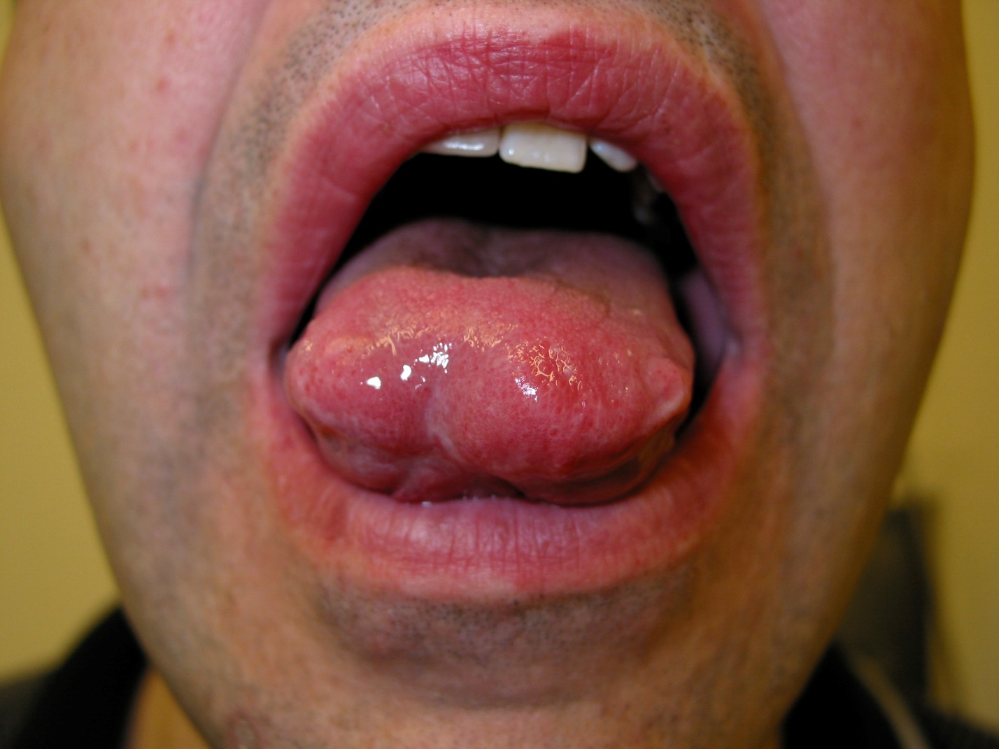
Approximately 90% of patients with sarcoidosis present with pulmonary manifestations (eg, dry cough, shortness of breath, chest pain), approximately 25% with cutaneous manifestations (eg, maculopapular eruption; erythematous scaling plaques; erythema nodosum; hypopigmentation or hyperpigmentation; scar or keloid), and approximately 13% with head and neck manifestations.5-8 In the head and neck region, the most commonly affected structures are the salivary glands and the cervical lymph nodes.1 The former may present as enlargement of the salivary glands and xerostomia and the latter as cervical lymphadenopathy (usually bilateral).2 Oral involvement is relatively rare and most often affects the buccal mucosa (30%), followed by the gingiva (20%), the tongue (16%), the lips (16%), and the palate (9%).9 Typically, oral sarcoidosis presents as one or more nontender firm swellings or nodules with normal overlying mucous membranes, as is illustrated in the Figure.7,9 At times, oral sarcoidosis presents as painless ulcers, gingivitis, gingival hyperplasia, or gingival recession.3,6
In the United States, the prevalence rate of sarcoidosis is 10 to 14 cases per 100,000 in the white population and 35.5 to 64 cases per 100,000 in the African American population.1,4,10 The age of onset peaks between 20 and 29 years and again between 45 and 65 years.2,3 The female to male ratio is 1.5 to 1.7,11
The exact etiology of sarcoidosis is not known. Presumably, the condition results from an exaggerated immune response to unknown environmental antigens in genetically susceptible individuals.12 The genetic predisposition is suggested by familial clustering, ethnic variations of disease prevalence, and increased concordance in monozygotic twins.1,5 Sarcoidosis is familial in 3.6% to 9.6% of cases, suggesting an autosomal recessive mode of inheritance with incomplete penetrance.12 Suffice to say, the majority of cases are sporadic.13
The diagnosis of sarcoidosis is mainly based on characteristic clinical features, typical radiologic features (bilateral hilar lymphadenopathy, pulmonary infiltrates), and histologic findings of noncaseating epithelial cell granulomas replete with Langerhans giant cells and a rim of lymphocytes.2,3,14 Supportive laboratory findings include elevation of acute-phase reactants such as erythrocyte sedimentation rate and C-reactive protein level, anemia, lymphocytopenia, elevated liver enzyme levels, and an increased serum angiotensin-converting enzyme level.4,10
Treatment depends on the extent and severity of the disease. No treatment is necessary for oral sarcoidosis apart from watchful observation in those with asymptomatic, mild, and focal disease, since spontaneous resolution is common.5,11 Topical and intralesional corticosteroids may be considered for limited or localized mucocutaneous involvement.5 Surgical excision is the treatment of choice for isolated, accessible, circumscribed, nodular lesions that are uncomfortable.11,13 For widespread and progressive lesions, systemic corticosteroids are the treatment of choice.13 For those who do not respond favorably to systemic corticosteroids, adjunct treatment with steroid-sparing drugs such as cyclosporine, methotrexate, cyclophosphamide, azathioprine, chlorambucil, pentoxifylline, infliximab, adalimumab, chloroquine, and hydroxychloroquine may be considered.3,10,12
REFERENCES:
- Al-Azri AR, Logan RM, Goss AN. Oral lesion as the first clinical presentation in sarcoidosis: a case report. Oman Med J. 2012;27(3):243-245.
- Radochová V, Radocha J, Laco J, Slezák R. Oral manifestation of sarcoidosis: a case report and review of the literature. J Indian Soc Periodontol. 2016;20(6):627-629.
- Bowers L, Brennan M. Oral complications of multiorgan disorders. Atlas Oral Maxillofac Surg Clin North Am. 2017;25(2):187-195.
- Dastoori M, Fedele S, Leao JC, Porter SR. Sarcoidosis—a clinically orientated review. J Oral Pathol Med. 2013;42(4):281-289.
- Alawi F. An update on granulomatous diseases of the oral tissues. Dent Clin North Am. 2013;57(4):657-671.
- Gill I, Siddiqi J. An oral lesion as the primary clinical manifestation of sarcoidosis. Ann R Coll Surg Engl. 2017;99(5):e135-e136.
- Gupta S, Tripathi AK, Kumar V, Saimbi CS. Sarcoidosis: oral and extra-oral manifestation. J Indian Soc Periodontol. 2015;19(5):582-585.
- Leung AKC, Leong KF, Lam JM. Erythema nodosum. World J Pediatr. 2018;14(6):548-554.
- Bouaziz A, Le Scanff J, Chapelon-Abric C, et al; Groupe Sarcoïdose Francophone. Oral involvement in sarcoidosis: report of 12 cases. QJM. 2012;105(8):755-767.
- Suresh L, Radfar L. Oral sarcoidosis: a review of literature. Oral Dis. 2005;11(3):138-145.
- Marcoval J, Mañá J. Specific (granulomatous) oral lesions of sarcoidosis: report of two cases. Med Oral Patol Oral Cir Bucal. 2010;15(3):e456-e458.
- Sankar V, Noujeim M. Oral manifestations of autoimmune and connective tissue disorders. Atlas Oral Maxillofac Surg Clin North Am. 2017;25(2):113-126.
- Motswaledi MH, Khammissa RA, Jadwat Y, Lemmer J, Feller L. Oral sarcoidosis: a case report and review of the literature. Aust Dent J. 2014;59(3):389-394.
- Tripathi P, Aggarwal J, Chopra D, Bagga S, Sethi K. Sarcoidosis presenting as isolated gingival enlargement: a rare case entity. J Clin Diagn Res. 2014;8(11):ZD25-ZD26.