
An Atlas of Lumps and Bumps, Part 30: Neurofibroma
Neurofibroma
Neurofibromas are benign peripheral nerve sheath tumors composed of an extracellular matrix and a heterogeneous mixture of Schwann cells, perineural cells, fibroblasts, and mast cells.1-3 Cutaneous neurofibromas (the most common type of neurofibroma) are the most common tumor of the peripheral nerve sheath.4They can develop as a solitary tumor or part of neurofibromatosis type 1 (NF1).5 Approximately 90% of neurofibromas occur sporadically while the rest are associated with NF1.1,5 The sex ratio is approximately equal.4 There is no ethnic or racial predilection.4Cutaneous neurofibromas are rarely seen in young children.1 They often appear during puberty or pregnancy, increase in size and number with age and are found in almost all adults with NF1.1 Clinically, cutaneous neurofibromas present as asymptomatic, soft (Fig. 1) or elastic, violaceous or skin-colored (Fig. 2), papules, nodules or pedunculated lesions (Fig. 3).1

Fig. 1. A neurofibroma is shown.

Fig. 2. Cutaneous neurofibromas present as asymptomatic, soft, or elastic.

Fig. 3. Neurofibromas can appear as violaceous or skin-colored papules, nodules, or pedunculated lesions.
The size of individual lesion is usually less than 2 cm in diameter.1 Cutaneous neurofibromas often invaginate into the skin and exhibit the "button-hole" sign (Fig. 4) when gentle digital pressure is applied to the surface and reappear on the release of pressure (Fig 5).1,3,4
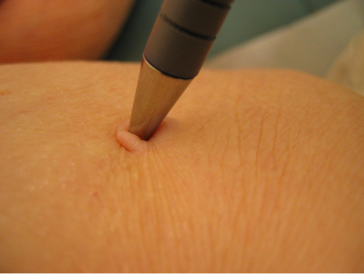
Fig. 4. Cutaneous neurofibromas can exhibit the "button-hole" sign.

Fig. 5. The size of individual lesion is usually less than 2 cm in diameter.
In contrast, subcutaneous neurofibromas, which are usually firm and painful, are deeply seated in the dermis and, therefore, less circumscribed.1 Plexiform neurofibromas result from diffuse thickening of nerve trunks and/or roots. They are found in 30 to 50% of individuals with NF1.2,6-8 Although plexiform neurofibromas are usually congenital, only approximately 44% of these tumors are detected by 5 years of age.6.9 Plexiform neurofibromas may become highly irregular, thick, and tortuous and feel like "a bag of worms".2,4,10 Lesions can be skin-colored or, more commonly, hyperpigmented (Fig. 6).4
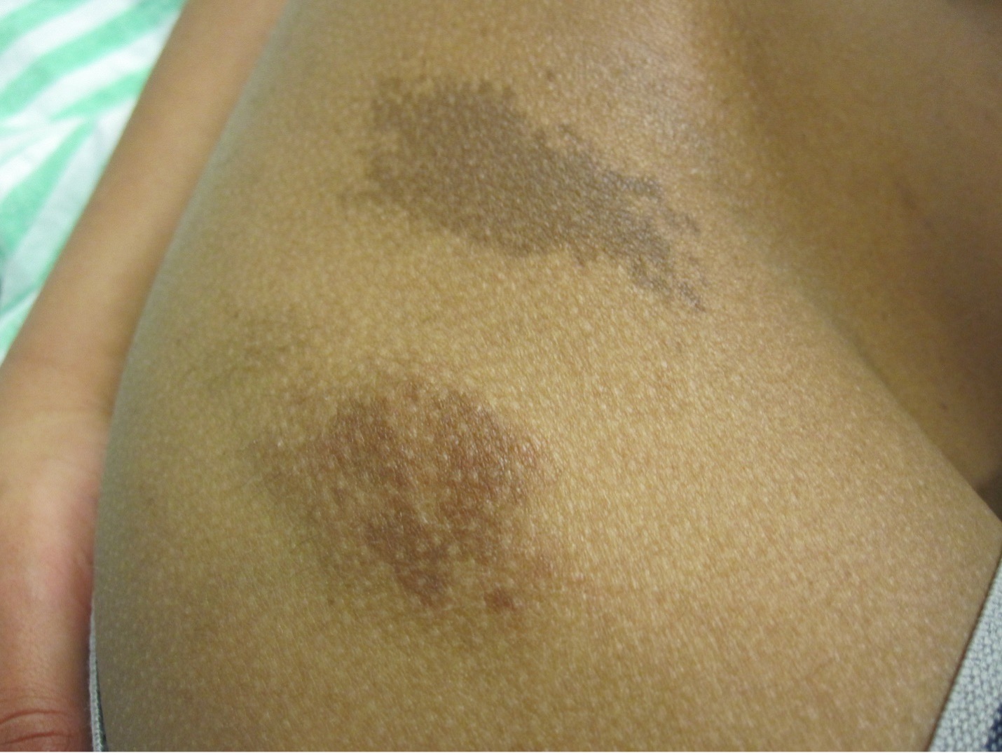
Fig. 6 Lesions can be skin-colored or, more commonly, hyperpigmented.
There may be overlying café-a-lait macules, hypertrichosis and, rarely, leukotrichia.7 Plexiform neurofibromas are slow-growing and usually asymptomatic.6,10They may be locally invasive and may present with pain, paresthesia, and numbness with involvement of adjacent structures.4 Plexiform neurofibromas are associated with an increased risk of bone erosion, spinal cord compression, carpal tunnel syndrome, overgrowth of an extremity, and malignant transformation.4,11 A diffuse neurofibroma, a rare variant of neurofibroma, most commonly involves the head and neck regions and has an onset mainly in adolescents and young adults.12
One of the diagnostic criteria for NF1 is two or more neurofibromas of any type or one plexiform neurofibroma.13 To make the diagnosis of NF1, two or more of the seven National Institutes of Health (NIH) diagnostic criteria must be present.13 In most children, the diagnosis of NF1 can be confidently made using the NIH diagnostic criteria by 8 years of age.14 Other diagnostic criteria include six or more café-au-lait spots, 1.5 cm or larger in postpubertal persons and 0.5 cm or larger in prepubertal persons; freckling in the axillary or inguinal regions; optic pathway glioma; two or more Lisch nodules; a distinctive osseous lesion, such as dysplasia of the sphenoid bone or dysplasia or thinning of the long bone cortex (with or without pseudoarthrosis); and a first-degree relative with NF1, according to the preceding criteria.13
AFFILIATIONS:
1Clinical Professor of Pediatrics, the University of Calgary, Calgary, Alberta, Canada
2Pediatric Consultant, the Alberta Children’s Hospital, Calgary, Alberta, Canada
3Dermatologist, Medical Director and Founder, the Toronto Dermatology Centre, Toronto, Ontario, Canada
4Associate Clinical Professor of Pediatrics, Dermatology and Skin Sciences, the University of British Columbia, Vancouver, British Columbia, Canada.
5Pediatric Dermatologist, the Pediatric Institute, Kuala Lumpur General Hospital, Kuala Lumpur, Malaysia
CITATION:
Leung AKC, Barankin B, Lam JM, Leong KF. An atlas of lumps and bumps, part 30: Neurofibromas. Consultant. 2023;63(8):e8. doi:10.25270/con.2023.08.000005.
CORRESPONDENCE:
Alexander K. C. Leung, MD, #200, 233 16th Ave NW, Calgary, AB T2M 0H5, Canada (aleung@ucalgary.ca)
EDITOR’S NOTE:
This article is part of a series describing and differentiating dermatologic lumps and bumps. To access previously published articles in the series, visit: https://www.consultant360.com/resource-center/atlas-lumps-and-bumps.
- Leung AKC, Barankin B, Leung AA. What are this woman’s widespread asymptomatic nodules? Neurofibromatosis type 1. Consultant. 2017;57:651-654.
- Anderson JL, Gutmann DH. Neurofibromatosis type 1. Handb Clin Neurol. 2015;132:75-86. doi: 10.1016/B978-0-444-62702-5.00004-4.
- Hernández-Martín A, Duat-Rodríguez A. An update on neurofibromatosis type 1: not just café-au-lait spots, freckling, and neurofibromas. An update. Part I. Dermatological clinical criteria diagnostic of the disease. Actas Dermosifiliogr. 2016;107(6):454-464. doi: 10.1016/j.ad.2016.01.004.
- Messersmith L, Krauland K. Neurofibroma. In: StatPearls [Internet]. Treasure Island (FL): StatPearls Publishing; 2020 Jan–2020 Aug 10. PMID: 30969529.
- Kim KS, Lee DG, Lee DH, Hwang JH, Lee SY. Slowly growing solitary neurofibroma of the thumb: A case report. Medicine (Baltimore). 2021;100(2):e23611. doi: 10.1097/MD.0000000000023611.
- Collins NC, Ayodeji EE, Motunrayo FO, Abayomi SB, Olufemi OI, Olusesan AL. Large penile plexiform neurofibroma in an 11-year old boy. Malawi Med J. 2018;30(1):49-51. doi: 10.4314/mmj.v30i1.11.
- Sahni MK, Chaurasia JK, Patra S. Circumscribed area of leukotrichia over plexiform neurofibroma: An important clue or mere coincidence? Indian Dermatol Online J. 2020;11(5):831-832. doi: 10.4103/idoj.IDOJ_606_19.
- Tchernev G, Chokoeva AA, Patterson JW, Bakardzhiev I, Wollina U, Tana C. Plexiform neurofibroma: A case report. Medicine (Baltimore). 2016;95(6):e2663. doi: 10.1097/MD.0000000000002663.
- Yocum AD, Bacharach D, Simon EL. An unusual presentation and treatment of a hemorrhaging plexiform neurofibroma. Am J Emerg Med. 2020:S0735-6757(20)30982-7. doi: 10.1016/j.ajem.2020.10.080.
- Poswal P, Bhutani N, Arora S, Kumar R. Plexiform neurofibroma with neurofibromatosis type I/ von Recklinghausen's disease: A rare case report. Ann Med Surg (Lond). 2020;57:346-350. doi: 10.1016/j.amsu.2020.08.015.
- Hu K, Ma H, Shen Y, Williams Z, Xu W. Plexiform neurofibroma as a cause of carpal tunnel syndrome in a radial deficiency patient. Br J Neurosurg. 2019;33(3):281-282. doi: 10.1080/02688697.2017.1333572.
- Nehete LS, Sharma R, Singh PR, Gupta S. Rapidly growing diffuse neurofibroma of the scalp with calvarial defect in a young woman: A rare entity. Surg Neurol Int. 2020;11:313. doi: 10.25259/SNI_213_2020.
- Gutmann DH, Aylsworth A, Carey JC, Korf B, Marks J, Pyeritz RE, Rubenstein A, et al. The diagnostic evaluation and multidisciplinary management of neurofibromatosis 1 and neurofibromatosis 2. JAMA. 1997;278(1):51-57. PMID: 9207339.
- DeBella K, Szudek J, Friedman JM. Use of the National Institutes of Health Criteria for diagnosis of neurofibromatosis 1 in children. Pediatrics. 2000;105(3):608-614. doi: 10.1542/peds.105.3.608.