
Peer Reviewed
A 29-Year-Old Woman With Unexplained Dyspnea
What's The "Take Home"?
Pearls From Clinical Cases
During the past 2 days, a 29-year-old woman has noted shortness of breath when walking and taking care of her 4-week-old daughter. She never experienced such symptoms previously.
HISTORY
The patient is otherwise healthy and takes no medications except for a multivitamin supplement with iron. Her pregnancy was uneventful, and she had a vaginal delivery 4 weeks ago. She has had no cough, fever, or hemoptysis.
PHYSICAL EXAMINATION
Blood pressure is 110/70 mm Hg; heart rate, 96 beats per minute; and respiration rate, 20 breaths per minute. The patient is afebrile. Neck veins are not distended. Breath sounds are normal; no wheezes or rales are audible. Heart rhythm is regular, without murmurs or gallops. She has 11 edema of the left ankle only.
LABORATORY AND IMAGING RESULTS
A D-dimer assay reveals a value of 2100 ng/mL. Cardiac troponin levels are normal. Ultrasonography demonstrates deep venous thrombosis in the left popliteal vein. Hemogram is normal. Creatinine level is 0.8 mg/dL.
Which of the following is the optimal therapy for this patient?
A. Insertion of a vena cava filter.
B. Initiation of anticoagulant therapy with subcutaneous low molecular weight heparin (LMWH).
C. Initiation of a short-course infusion of intravenous thrombolytic therapy.
D. Initiation of anticoagulant therapy with intravenous unfractionated heparin.
(Answer and discussion begins on next page.)
What's The "Take Home"?
CORRECT ANSWER: B
The findings in this case are diagnostic of acute pulmonary embolism. Although this disorder has a long history of diagnostic conundrums, paradigms, and techniques, more than sufficient data are present here to support the diagnosis.
Clinical findings. The patient exhibits the classic symptom of new and unexplained dyspnea. There is no other obvious or, for that matter, subtle cause. In addition, the risk of pulmonary embolism increases during the 6-week period after delivery. Her D-dimer value is very high (although her postpartum state probably also contributes to the elevation), and ultrasonography confirmed the presence of a deep venous thrombosis. Thus, multidetector CT is not necessary for this patient; in fact, there is at least a theoretical contraindication of significant radiation exposure.
Treatment. The issue here is how to manage the pulmonary embolism. The clinical findings demonstrate that she is hemodynamically stable. Her blood pressure is normal and, although not specifically further confirmed by detailed right ventricular testing (eg, measurement of B-type natriuretic peptide, echocardiography),1 her cardiac status is good. She can thus be considered at low risk, without evidence of right ventricular injury or dysfunction.1 These findings then predicate the most appropriate line of therapy.
Vena cava filters (choice A) have an extremely focal and limited indication in acute pulmonary embolism; they should be reserved for patients with contraindications to anticoagulants.2,3 These contraindications include current active bleeding or very recent bleeding that has now ceased.
 More of the allegedly retrievable filters are placed than are actually retrieved.1,2 It must be emphasized that filters have brief and marginal, at best, benefit compared with anticoagulation, and they should not be used in patients without contraindications to the latter.4 And, since the filters themselves predispose to subsequent venous thrombosis in the legs, any patient in whom a filter has been placed should receive a course of anticoagulant therapy once the risk of bleeding is past.1,2
Thrombolytic therapy (choice C) is another very aggressive tactic used in hemodynamically unstable patients (eg, those with shock or hypotension) and in patients who have evidence of right ventricular injury (eg, elevated troponin levels or right ventricular hypokinesis/dilatation). In such instances, a 2-hour infusion of a thrombolytic agent has been shown to result in more rapid resolution without mortality benefit in the latter and actual reduction in mortality in the former.5,6 This patient does not exhibit any of the findings that would indicate the need for this therapy.
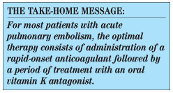
Thus, administration of a rapid-onset anticoagulant followed by a period of therapy with an oral vitamin K antagonist is the optimal treatment here, as it is in the vast majority of patients with acute pulmonary embolism. The agents available are the traditional anticoagulants, unfractionated heparin and LMWH, and the newer pentasaccharide agent, fondaparinux. Both LMWHs and fondaparinux have far more convenient pharmacology, which allows for subcutaneous administration without the cumbersome intravenous administration and partial thromboplastin time monitoring
of unfractionated heparin. As a result, these agents are preferred by most patients and physicians.
Cost can also be a factor, depending on hospital and third-party payor policy. An important consideration is the renal clearance of LMWH and fondaparinux. These drugs accumulate in patients with renal failure (creatinine clearance of less than 30 mL/min); in such cases, there is no choice but to use unfractionated heparin. Otherwise, overwhelming data have demonstrated that all these anticoagulants are equivalent in efficacy and safety. Therefore, LMWH (choice B) is preferable to unfractionated heparin (choice D) because of ease of use.
Outcome of this case. A LMWH (enoxaparin) was initiated in the therapeutic dosage of 1 mg/kg twice daily by subcutaneous route. By the morning of the second day, the patient’s symptoms had markedly improved. On day 3 she was discharged, and the LMWH was continued. Warfarin was initiated, and the plan was to discontinue the LMWH once the INR target of 2.0 to 3.0 was attained, but with the assurance that a minimum of 5 days of LMWH be given.
Since the pulmonary embolism occurred in the setting of a reversible, documented risk factor (postpartum state), anticoagulation will be continued for 3 to 6 months. A workup for the presence of a hereditary hypercoagulable state will be done after the warfarin is discontinued. ■
References
1. Agnelli G, Becattini C. Acute pulmonary embolism. N Engl J Med. 2010;363:266-274.
2. Kearon C, Kahn SR, Agnelli G, et al. Antithrombotic therapy for venous thromboembolic disease:American College of Chest Physicians Evidence-Based Clinical Practice Guidelines (8th edition).Chest. 2008;133(suppl):454S-545S.
3. Torbicki A, Perrier A, Konstantinides S, et al. Guidelines on the diagnosis and management of acute pulmonary embolism: the Task Force for the Diagnosis and Management of Acute Pulmonary Embolism of the European society of Cardiology (ESC). Eur Heart J. 2008;29:2276-2315.
4. Decousus H, Leizorovicz A, Parent F, et al. A clinical trial of vena caval filters in the prevention of pulmonary embolism inpatients with proximal deep-vein thrombosis. Prévention du Risque d’Embolie Pulmonaire par Interruption Cave Study Group. N Engl J Med. 1998;338:409-415.
5. Konstantinides S, Geibel A, Heusel G, et al. Heparin plus alteplase compared with heparin alone in patients with submassive pulmonary embolism. N Engl J Med. 2002;347:1143-1150.
6. Wan S, Quinlan DJ, Agnelli G, Eikelboom JW. Thrombolysis compared with heparin for the initial treatment of pulmonary embolism: a meta-analysis of the randomized controlled trials. Circulation.2004;110:744-749.