
Peer Reviewed
What’s the Best Approach to Diagnosis and Treatment of This Man’s Scalp Lesions?
Authors:
Donna Gensheimer, FNP-BC; Gail O’Toole, ANP-BC; and Kellie Murphy-Roche, RN, BSN
Citation:
Gensheimer D, O'Toole G, Murphy-Roche K. What’s the best approach to diagnosis and treatment of this man’s scalp lesions? Consultant. 2017;57(6):362.
An 82-year-old man presented with a 6-year history of scalp lesions, which consisted of recurrent pustules, thick yellow-brown crusts, and inflamed eroded plaques overlying a healed graft site. The lesions were not pruritic or painful, but the appearance of the erosions and crusts were embarrassing to the patient, and he wore a hat to cover the area.
The patient’s history included squamous cell carcinoma of the scalp in 2005, for which he had undergone skin grafting. His dermatologic history also included basal cell carcinoma and actinic keratosis. Recent treatments with debridement, topical and oral antibiotics, and cryotherapy (thermal destruction with liquid nitrogen) had all been ineffective.

Answer on next page
Answer: Erosive pustular dermatosis
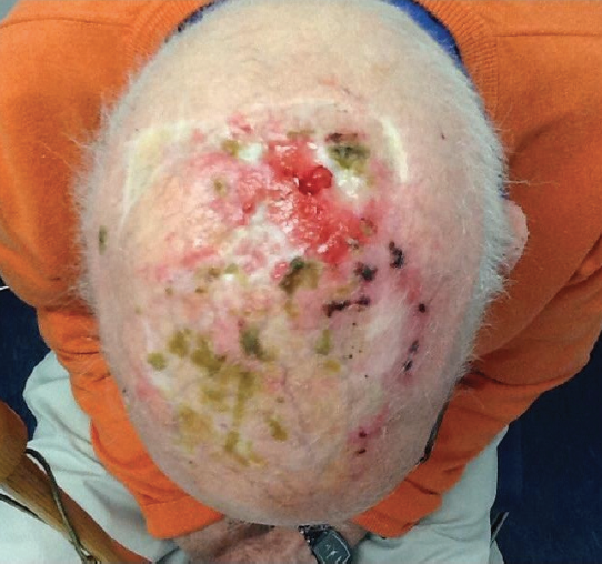
An 82-year-old man presented with a 6-year history of scalp lesions, which consisted of recurrent pustules, thick yellow-brown crusts, and inflamed eroded plaques overlying a healed graft site (Figure 1). The lesions were not pruritic or painful, but the appearance of the erosions and crusts were embarrassing to the patient, and he wore a hat to cover the area.
The patient’s history included squamous cell carcinoma (SCC) of the scalp in 2005, for which he had undergone skin grafting. His dermatologic history also included basal cell carcinoma and actinic keratosis. Recent treatments with debridement, topical and oral antibiotics, and cryotherapy (thermal destruction with liquid nitrogen) had all been ineffective.
Repeated culture tests had been done over the years, the results of which had been positive for heavy growth of Staphylococcus aureus; accordingly, the patient had been treated with topical and oral antibiotics, which had led to only temporary improvement.
Results of the most recent biopsy of the lesions demonstrated granulation tissue and scar tissue, consistent with resolving ruptured cysts or follicles, and no evidence of basal cell carcinoma or SCC. The most recent culture test results showed moderate growth of S aureus.
Based on the results of biopsy and bacterial culture testing, the man received a diagnosis of erosive pustular dermatosis (EPD).
Discussion
EPD is a relatively rare or perhaps underdiagnosed condition. It is a noninfectious inflammatory disorder that develops on the scalp and/or the lower extremities. EPD is mainly reported in the elderly population,1 and it is a chronic dermatosis of unknown etiology.1,2 Causes can include chronic sun exposure, trauma (mechanical and chemical), and skin grafting.
The lack of specific histologic features can make the diagnosis of EPD difficult. Histologic features are nonspecific changes of chronic inflammation, and fibrosis of the dermis with adnexal destruction also may be seen.3 Treatment can be difficult, and recurrence is common.
EPD can lead to scarring alopecia. The disorder requires ongoing management and monitoring given that patients with it are at increased risk of developing nonmelanoma skin cancer.3
No treatment algorithm exists for EPD, but high-potency topical corticosteroids have been used safely and with good results.5 Other reportedly successful treatment options include tacrolimus ointment, 0.1%; calcitriol cream, 0.005%; and dapsone gel, 5%.4,5 In addition, acitretin and anti-inflammatory antibiotics such as minocycline can be used but with limited reported success.5
Other possible diagnoses to consider in cases of EPD are SCC, pseudomonal cellulitis, and tinea capitis.
SCC is a malignant tumor of the epithelial keratinocytes. SCC is common in the skin of sun-exposed areas.6 Its development is associated with chronic sun exposure (such as in persons who work outdoors), sunburns, exposure to ionizing radiation, and having light skin. It can arise in areas of previous burns or scar tissue.
Cellulitis resulting from infection with Pseudomonas aeruginosa can present as a localized infection characterized by erosions, tissue necrosis, and greenish purulent material. Pseudomonal cellulitis can be associated with pain, and an odor may be present along with satellite lesions of pustules and vesicles.7
Tinea capitis is a dermatophytic trichomycosis of the scalp. It can present with scaly, patchy alopecia, as well as pustules, papules, and pruritus. Tinea capitis is more often seen in children, with 90% of cases caused by Trichophyton tonsurans. Inflammation can be mild or severe and can subsequently lead to a secondary infection.8
Outcome of the Case
Our initial treatment plan was the application of diluted vinegar compresses daily to help remove crusts; a high-potency topical corticosteroid ointment, applied twice daily for 2 weeks, to address inflammation; and oral cephalexin, 500 mg 3 times a day for 7 days, to treat the S aureus infection.
At the 3-week follow-up visit, the patient’s scalp had cleared (Figure 2). He was instructed to decrease the high-potency topical corticosteroid applications to once a day on weekends only for 4 more weeks. He also was instructed to discontinue the vinegar compresses and to follow up for a recheck in 4 to 5 weeks.
Five weeks later, the patient's scalp lesions were recurrent, now with purulent discharge and odor (Figure 3). Results of culture test were negative for bacteria. He was instructed to restart the vinegar compresses to help remove the crusts; to cleanse the area daily with a mild unscented soap; to continue the high-potency topical corticosteroid applications once a day on weekends only; and to follow up in 3 to 4 weeks for a recheck.
At a follow-up visit 3 weeks later, the patient’s scalp had healed, with no open areas or crusting (Figure 4). He was instructed to discontinue the vinegar soaks, but to continue the daily cleansing with mild soap. He also was instructed to continue applying the corticosteroid ointment once a day only on weekends, but if the lesions worsened, to increase the applications to once every day.
At a follow-up visit 4 weeks later, the patient’s scalp remained clear, and he was to schedule to be reassessed every 3 months thereafter.
LISTEN TO THE PODCAST WITH DONNA GENSHEIMER HERE.
Donna Gensheimer, FNP-BC, is a nurse practitioner at South Coast Dermatology and Cosmetic Center in Weymouth, Massachusetts.
Gail O’Toole, ANP-BC, is a nurse practitioner formerly at South Coast Dermatology and Cosmetic Center in Weymouth, Massachusetts.
Kellie Murphy-Roche, RN, BSN, is a nurse at Boston Medical Center in Boston, Massachusetts, and for the Town of Scituate, Massachusetts.
REFERENCES:
- Stan TR, Starace M, Bruni F, Misciali C, Piraccini BM. Erosive pustular dermatitis of the scalp: case series. Clin Dermatol. 2014;2(1):59-63.
- Laffitte E, Kaya G, Piguet V, Saurat J-H. Erosive pustular dermatosis of the scalp: treatment with topical tacrolimus. Arch Dermatol. 2003;139(6):712-714.
- Corradin MT, Forcione M, Guilioni E, Fiorentino R, Ferrazzi A, Alaibac M. Erosive pustular dermatosis of the scalp induced by imiquimod. Case Rep Dermatol Med. 2012;2012:828749.
- Broussard KC, Berger TG, Rosenblum M, Murase JE. Erosive pustular dermatosis of the scalp: A review with a focus on dapsone therapy. J Am Acad Dermatol. 2012;66(4)680-686.
- Semkova K, Tchernev, Wollina U. Erosive pustular dermatosis (chronic atrophic dermatosis of the scalp and extremities). Clin Cosmet Investig Dermatol. 2013;6:177-182.
- Wolff K, Johnson RA, Saavedra AP, Roh EK. Squamous cell carcinoma in situ. In: Wolff K, Johnson RA, Saavedra AP, Roh EK. Fitzpatrick’s Color Atlas and Synopsis of Clinical Dermatology. 8th ed. New York, NY: McGraw-Hill; 2017:227-230.
- Habif TP. Pseudomonas aeruginosa infection. In: Habif TP. Clinical Dermatology: A Color Guide to Diagnosis and Therapy. 5th ed. Philadelphia, PA: Mosby Elsevier; 2010:363-371.
- Habif TP. Dermatophyte fungal infections. In: Habif TP. Clinical Dermatology: A Color Guide to Diagnosis and Therapy. 5th ed. Philadelphia, PA: Mosby Elsevier; 2010:491-523.