
Peer Reviewed
What Are These Asymptomatic Papules on a Man’s Cheeks and Forehead?
Authors:
Alexander K. C. Leung, MD, and Benjamin Barankin, MD
Citation:
Leung AKC, Barankin B. What are these asymptomatic papules on a man’s cheeks and forehead? Consultant. 2017;57(6):355-357.
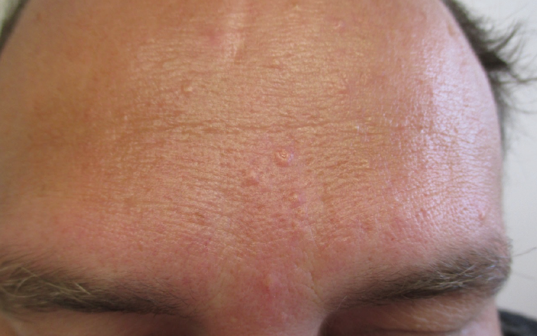
A 40-year-old white man presented with multiple flesh-colored lesions on the forehead that he had first noticed a year or so ago. The lesions were asymptomatic. There had been no discharge or secretion from the lesions. The lesions had increased slowly in size and number over time.
The patient was otherwise healthy and was not taking any medication. There was no family history of similar skin lesions. He had a history of an oily complexion.
On examination, multiple, flesh-colored, firm, oval, papules with sharply demarcated borders were noted on the forehead and cheeks. The papules measured 2 to 3 mm in diameter. The surface of the papules was smooth and shiny. Some of them had central umbilication. No other cutaneous or systemic findings were noted.

Answer on next page
Answer: Sebaceous hyperplasia
Dermoscopy results showed well-defined, oval and asymmetric, milky white, cloudlike structures. Skin biopsy results showed multiple enlarged well-defined sebaceous glands with numerous mature sebaceous lobules grouped in a grapelike pattern discharging into their individual centrally located wide sebaceous ducts.
A diagnosis of sebaceous hyperplasia was made. The patient’s dermatologist carefully electrodesiccated the lesions with very nice results and no scarring.
EPIDEMIOLOGY
Sebaceous hyperplasia is a common and benign proliferation of sebaceous glands that affects an estimated 1% of the general population.1 Although sebaceous hyperplasia can occur in individuals of all races, it is more commonly observed in the white population.2 The condition is seen mainly in middle-aged and elderly individuals, with a slight male predominance.1,3 Premature sebaceous hyperplasia, on the other hand, occurs mainly during puberty or soon after, without sex predominance.1
ETIOPATHOGENESIS
The exact etiology is not known. Many authors believe that a decrease in androgen levels with advancing age leads to a decrease in cellular turnover rate of sebocytes.3 This process in turn leads to an increase in the number of and thus crowding of sebocytes within the sebaceous gland, with resultant sebaceous hyperplasia.3 A recent study, however, showed that no significant changes occur in circulating androgen levels in patients with sebaceous hyperplasia.2 Future studies are necessary to confirm or refute this finding.
Besides androgens, insulin and epidermal growth factor may also have an operative role.4 Other predisposing factors include chronic sun exposure, immunosuppression (in particular, organ transplant recipients receiving cyclosporine), and hemodialysis.1 There is also a genetic predisposition. Familial cases with an autosomal dominant mode of inheritance with incomplete penetrance have been reported.5 Studies have shown that deletion of PRDM1 (also known as BLIMP1) may result in sebaceous gland hyperplasia.6 Individuals with Muir-Torre syndrome (an autosomal recessive disorder with sebaceous neoplasm, keratoacanthoma, and internal malignancy), dermatofibroma, pachydermatosis, or X-chromosomal hypohidrotic ectodermal dysplasia (XHED) are at risk for sebaceous hyperplasia.7
HISTOPATHOLOGY
Histologic examination of a classic lesion shows increased size and number of mature sebaceous lobules opening into a dilated central sebaceous duct, appearing like a bunch of grapes.3 The ductal opening in the epidermis corresponds to the central umbilication.
CLINICAL MANIFESTATIONS
Sebaceous hyperplasia most often presents as a solitary, or more commonly, as multiple discrete, yellow or flesh-colored, dome-shaped papules in an area where sebaceous glands are abundant.1 Some of the lesions have central umbilication.1 Individual lesions usually measure 2 to 5 mm in diameter. The lesions are often asymptomatic.
The face, in particular the forehead, cheeks, and nose, is most commonly affected.3 Other less common sites of involvement include the neck, chest, areola, nipple, penis, scrotum, vulva, and buccal mucosa. It has been the authors’ experience that patients often have a history of having an oily complexion.
While the majority of cases are sporadic and occur at an older age, premature sebaceous hyperplasia with an earlier age of onset occurs in familial cases and individuals with Muir-Torre syndrome, pachydermatosis, and XHED.3,7
DIAGNOSIS and Differential DIagnosis
The diagnosis is usually clinical and can be aided by dermoscopy. Dermoscopy findings of a typical lesion show central umbilication surrounded by a well-defined, milky-white, cloudlike structure (“cumulus sign”8 or “bonbon toffee sign”9). Arborizing blood vessels (blood vessels with multiple treelike branches) and nonarborizing and wreathlike blood vessels at the periphery of the lesion (“crown vessels”) can also be seen.8,9 Sometimes the ostium of the sebaceous gland is visible as a small crater. Examination findings using reflectance-mode confocal microscopy show enlarged sebaceous lobules consisting of cuboidal cells with centrally located nuclei and a dilated sebaceous duct.10 A skin biopsy should be considered if the diagnosis is in doubt, since basal cell carcinoma can occasionally mimic the lesions of sebaceous hyperplasia.
Sebaceous hyperplasia should be differentiated from sebaceous nevus, sebaceous adenoma, sebaceous epithelioma, basal cell carcinoma, sebaceous carcinoma, molluscum contagiosum, milia en plaque, and xanthoma.7
Treatment
Although the lesions are cosmetically unsightly and socially embarrassing for some patients, the condition is benign, and treatment is mainly for cosmesis.
When treatment is desired, the options include cryotherapy, electrodesiccation, curettage, topical bichloracetic or trichloroacetic acid, shave excision, oral isotretinoin, lasers, intense pulsed light, and topical 5-aminolevulinic acid with photodynamic therapy.3,7
Consider referral to a dermatologist for diagnostic confirmation or treatment.
Alexander K. C. Leung, MD, is clinical professor of pediatrics at the University of Calgary and a pediatric consultant at the Alberta Children’s Hospital in Calgary, Alberta, Canada.
Benjamin Barankin, MD, is a dermatologist and the medical director and founder of the Toronto Dermatology Centre in Toronto, Ontario, Canada.
REFERENCES:
- Wang Q, Liu J-M, Zhang Y-Z. Premature sebaceous hyperplasia in an adolescent boy. Pediatr Dermatol. 2011;28(2):198-200.
- Tagliolatto S, Alchorne MM, Enokihara M. Sebaceous hyperplasia: a pilot study to correlate this skin disease with circulating androgen levels. An Bras Dermatol. 2011;86(5):917-923.
- Yu C, Shahsavari M, Stevens G, Liskanich R, Horowitz D. Isotretinoin as monotherapy for sebaceous hyperplasia. J Drugs Dermatol. 2010;9(6):699-701.
- Tóth BI, Oláh A, Szöllősi AG, Czifra G, Bíró T. “Sebocytes makeup”: novel mechanisms and concepts in the physiology of the human sebaceous glands. Pflugers Arch. 2011;461(6):593-606.
- Boonchai W, Leenutaphong V. Familial presenile sebaceous gland hyperplasia. J Am Acad Dermatol. 1997;36(1):120-122.
- Horsley V, O’Carroll D, Tooze R, et al. Blimp1 defines a progenitor population that governs cellular input to the sebaceous gland. Cell. 2006;126(3):597-609.
- Richey DF. Aminolevulinic acid photodynamic therapy for sebaceous gland hyperplasia. Dermatol Clin. 2007;25(1):59-65.
- Bryden AM, Dawe RS, Fleming C. Dermatoscopic features of benign sebaceous proliferation. Clin Exp Dermatol. 2004;29(6):676-677.
- Oztas P, Polat M, Oztas M, et al. Bonbon toffee sign: a new dermatoscopic feature for sebaceous hyperplasia. J Eur Acad Dermatol Venereol. 2008;22(10):1200-1202.
- Propperova I, Langley RG. Reflectance-mode confocal microscopy for the diagnosis of sebaceous hyperplasia in vivo. Arch Dermatol. 2007;143(1):134.