
Peer Reviewed
Subungual Exostosis: A Bony Tumor Often Misdiagnosed as a Wart
Authors:
Vienna G. Katana, DO, and John E. Jackson, MD
Citation:
Katana VG, Jackson JE. Subungual exostosis: a bony tumor often misdiagnosed as a wart. Consultant. 2017;57(8):490-492.
A 5-year-old girl presented with a 2-month history of a rapidly growing and painful “lump” on her right fourth toe. There had been no known associated trauma, and the child complained of pain while wearing closed-toe shoes.
The patient’s treatment course had been complicated by a 1-month home application of topical over-the-counter salicylic acid and a single trial of cryotherapy performed by her primary care physician. The patient had been referred to our dermatology department.
On physical examination at her initial dermatology visit, a large subungual blister was present, obscuring the underlying lesion; therefore, the patient was directed to return to the clinic in 3 weeks, after the blister had resolved, to allow a better assessment of the primary lesion.
Evaluation at her visit 3 weeks later showed resolution of the blister, but with a hard, white, hyperkeratotic, 8-mm nodule extending from the distomedial portion of the right fourth toe. The associated nail plate was displaced laterally (Figure 1). A subungual exostosis (SE) was suspected clinically and confirmed radiographically (Figure 2).

Figure 1. A hyperkeratotic nodule extended from the distomedial portion of the right fourth toe.
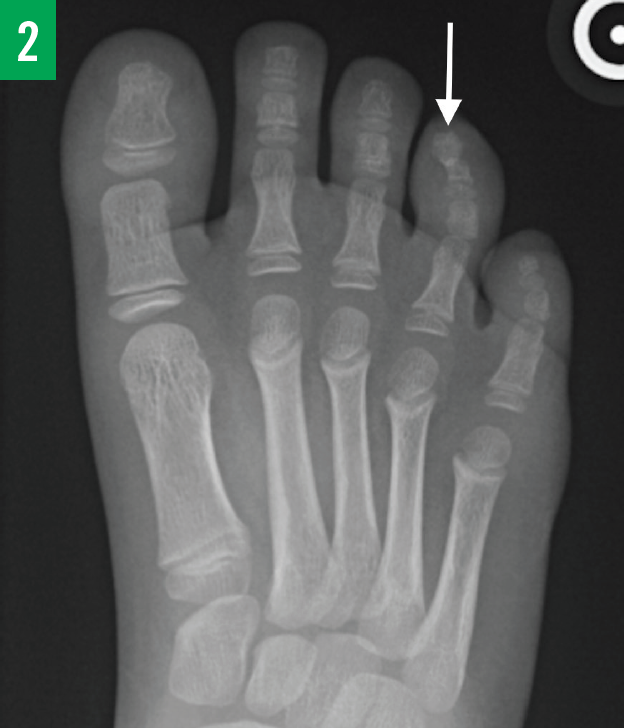
Figure 2. Anteroposterior radiograph demonstrating a bony outgrowth arising from the tuft of the right fourth distal phalanx.
The patient later underwent surgical excision of the bony growth utilizing a horizontal or fish-mouth incision at the margin of the sterile nail matrix and plantar skin. The residual hyperkeratotic mass was also resected from the overlying matrix/nail bed. Histologic examination of a specimen showed bony trabeculae surrounded by a fibrous proliferation with a regular maturation pattern.
The girl was seen 3 weeks postoperatively, at which time a scab was noted on the nail bed, in addition to remnants of the old nail plate, preserving an open nail fold. Her plan of care outlined a 3- to 4-week follow-up to evaluate the integrity of the nail and monitor for associated complications. We presumed an uneventful recovery, since the patient subsequently was lost to follow-up.
Discussion
SEs are solitary, benign osseous tumors of the toes or fingers.1 Although evidence on the exact cause is inconclusive, some authors have hypothesized that the condition results from reactive metaplasia of the fibrocartilaginous tuft of the distal phalanx due to chronic irritation and microtrauma.1 Neoplastic processes have been described, with a reproducible translocation t(X;6)(q22;q13-14) linked to SEs.1,2
Epidemiology
Relatively rare conditions such as SE are often characterized by single case reports, retrospective case series, and retrospective reviews. Moreover, this entity has been described as a “fringe condition,” whereby it is encountered clinically by many specialties, thus limiting the ability to gather, digest, and assimilate meaningful information about its epidemiology and treatment.3 In correlation with epidemiologic features described by systematic reviews in the 1990s, DaCambra and colleagues found that the average age of onset is 26 years, with the hallux being the site of tumor growth in 80% of cases.1 In contrast with the demographics described in older literature,4 DaCambra and colleagues also reported a much higher percentage of SEs occurring in the pediatric population (55% vs the prior report of 16%), and they reported that the sexes appear to be equally affected (1:1 vs the prior report of a 2:1 female-to-male ratio).
Clinical Manifestations
Although a patient’s clinical presentation can vary given the extent of overlying skin and nail deformity, common manifestations of SE include pain, erythema, and nail deformity evolving over several months,5 often leading to discomfort while wearing shoes.
Diagnosis and Differential Diagnosis
Subungual masses can often masquerade as refractory warts in the primary and specialty care setting.6-9 A retrospective study by Malkoc and colleagues revealed that approximately 25% of patients with SE referred to one institution had been inappropriately treated for verruca.10 Classic features of verruca vulgaris include hyperkeratosis that distorts normal skin lines, brown or red dots that represent thrombosed capillary loops, and punctate bleeding when the surface of the lesion is pared away.11,12 These discerning features may or may not have been considered in the cases described by Malkoc and colleagues.
Other soft-tissue tumors to consider are squamous cell carcinoma, glomus tumor, pyogenic granulomas, inclusion cyst, and malignant melanoma.11,13 Although this broad differential is believed to delay diagnosis in up to 10% of cases,1 these other soft-tissue tumors lack the osseous component of SE visualized on plain radiographic imaging.13 Moreover, certain diagnoses are less likely to present in the adolescent population.
Subungual osteochondroma has a similar osseous component to SE. Although the features of the 2 conditions are clinically and radiologically alike, histologically, osteochondromas form from endochondral ossification and have a cartilaginous cap made of hyaline. In contrast, the SE bony tumor is formed from fibrous tissue with a fibrocartilaginous cap.1,14 SE occurs in isolation, whereas osteochondromatosis (multiple osteochondromas), also known as hereditary multiple exostoses, has been described and classified as an autosomal dominant genetic disorder.15
While there have been no reports of malignant degeneration of an SE, Valero and colleagues reported an isolated case of a subungual squamous cell carcinoma arising at the site of a longstanding SE.16 In contrast, single osteochondromas and multiple osteochondromas have an established but rare malignant transformation rate of less than 1%17 and 0.5% to 5%,15 respectively. Despite these similarities and differences, the true clinical relevance of discerning a subungual SE from an isolated osteochondroma is unknown.5
Treatment and Prognosis
Surgical treatment approaches are diverse, without clear evidence to show a superior technique.1 Marginal excision using the principle of curetting or burring down to normal trabecular bone, while preserving the nail bed and matrix, is the standard of care. Two common approaches include the dorsal excision and the horizontal fish-mouth excision. The dorsal approach has been indicated for lesions that protrude from and thus damage the nail bed. The distorted nail would be removed, with attempts to repair the underlying nail bed once the bony tumor has been completed resected. In contrast, the fish-mouth approach spares the resident nail and nail bed, predicting rapid recovery and excellent cosmesis; however, higher recurrence rates have been reported.18
There is a tedious balance given that incomplete excision is the suspect for recurrence (estimated at 4%), while an overly aggressive dissection may complicate recovery and lead to higher rates of onychodystrophy (complication rates estimated at 16%).1 Some authors suggest preoperative evaluation and planning utilizing magnetic resonance imaging to detect the degree to which the tumor abuts the nail bed and matrix.19 Postoperative wound management remains a challenge; techniques using vacuum-assisted closure to mitigate these complications have been described.5
The Take-Home Message
Subungual tumors will be encountered by a diverse group of medical providers. Clinicians should play close attention to the subtle features of verrucous lesions and other exophytic tumors that can masquerade as SE. A simple, noninvasive evaluation with radiography to reveal the osseous component will prevent unnecessary and inappropriate treatment. There is currently low-level evidence for best practices; therefore, future studies are needed to enhance evidence-based recommendations.
Vienna G. Katana, DO, is at the Surface Warfare Medical Institute, a detachment of the Navy Medicine Operational Training Center, in San Diego, California.
John E. Jackson, MD, is at the Naval Medical Center San Diego Department of Dermatology in San Diego, California.
Disclaimer: The views expressed in this article are those of the authors and do not reflect the official policy or position of the Department of the Navy, the Department of Defense, or the US Government.
REFERENCES:
- DaCambra MP, Gupta SK, Ferri-de-Barros F. Subungual exostosis of the toes: a systematic review. Clin Orthop Relat Res. 2014;472(4):1251-1259.
- Mertens F, Möller E, Mandahl N, et al. The t(X;6) in subungual exostosis results in transcriptional deregulation of the gene for insulin receptor substrate 4. Int J Cancer. 2011;128(2):487-491.
- Damron TA. CORR Insights®: subungual exostosis of the toes: a systematic review. Clin Orthop Relat Res. 2014;472(4):1260-1261.
- Davis DA, Cohen PR. Subungual exostosis: case report and review of the literature. Pediatr Dermatol. 1996;13(3):212-218.
- DaCambra MP, Gupta SK, Ferri-de-Barros F. A novel management strategy for subungual exostosis. BMJ Case Rep. 2013;2013. doi:10.1136/bcr-2013-200396
- Daragad MS, Srinivas SD, Varghese J. Exostosis masquerading as a subungual wart. Indian Dermatol Online J. 2014;5(1):92-93.
- Bach DQ, McQueen AA, Lio PA. A refractory wart? Subungual exostosis. Ann Emerg Med. 2011;58(5):e3-e4.
- Campanelli A, Borradori L. Subungual exostosis. N Engl J Med. 2008;359(25):e31.
- Glick SR. Subungual osteochondroma of the third toe. Consult Pediatrician. 2013;12(9):422-424.
- Malkoc M, Korkmaz O, Keskinbora M, et al. Surgical treatment of nail bed subungual exostosis. Singapore Med J. 2016;57(11):630-633.
- Perez M, Engel G. Enlarging, painful nodule under the toenail. Am Fam Physician. 2014;89(10):793-794.
- Mulhem E, Pinelis S. Treatment of nongenital cutaneous warts. Am Fam Physician. 2011;84(3):288-293.
- Ward CM, Dittmer A. Subungual exotosis of the finger: case report and review of the literature. Iowa Orthop J. 2013;33:228-231.
- Lee SK, Jung MS, Lee YH, Gong HS, Kim JK, Baek GH. Two distinctive subungual pathologies: subungual exostosis and subungual osteochondroma. Foot Ankle Int. 2007;28(5):595-601.
- Mărginean CO, Meliţ LE, Mărginean MO. Daughter and mother diagnosed with hereditary multiple exostoses: a case report and a review of the literature. Medicine (Baltimore). 2017;96(1):e5824.
- Valero J, Gallart J, Gonzalez D, Deus J, Lahoz M. Subungual squamous cell carcinoma and exostosis in third toe—case report and literature review. J Eur Acad Dermatol Venereol. 2014;28(10):1292-1297.
- Bernard SA, Murphey MD, Flemming DJ, Kransdorf MJ. Improved differentiation of benign osteochondromas from secondary chondrosarcomas with standardized measurement of cartilage cap at CT and MR imaging. Radiology. 2010;255(3):857-865.
- Başar H, İnanmaz,ME, Başar B, Bal E, Köse KÇ. Protruded and nonprotruded subungual exostosis: differences in surgical approach. Indian J Orthop. 2014;48(1):49-52.
- Higuchi K, Oiso N, Yoshida M, Kawada A. Preoperative assessment using magnetic resonance imaging for subungual exostosis beneath the proximal region of the nail plate. Case Rep Dermatol. 2011;3(2):155-157.