
Peer Reviewed
Hyperpigmented Patches on a Woman’s Sun-Exposed Face: What’s the Cause?
Authors:
Alexander K. C. Leung, MD, and Benjamin Barankin, MD
Citation:
Keung AKC, Barankin B. Hyperpigmented patches on a woman’s sun-exposed face: what’s the cause? Consultant. 2017;57(8):485-489.
A 45-year-old woman presented with brownish macules and patches on her face. The pigmentation had developed approximately 10 years ago with the birth of her third child. The lesions had gradually increased in size and number over time. She had taken oral contraceptives for a few years after the birth of her second child. She enjoyed a variety of outdoor activities, and she reported that the pigmentation was more pronounced with sun exposure.
Her past health was unremarkable, and she was not on any topical or oral medications. She denied any history of inflammatory dermatosis prior to the appearance of her skin problem. The family history was not contributory.
Physical examination revealed symmetric, well-circumscribed, brownish macules and patches with irregular borders on the forehead, cheeks, nose, upper lip, and chin. The rest of the physical examination findings were normal.

Answer on next page
Answer: Melasma
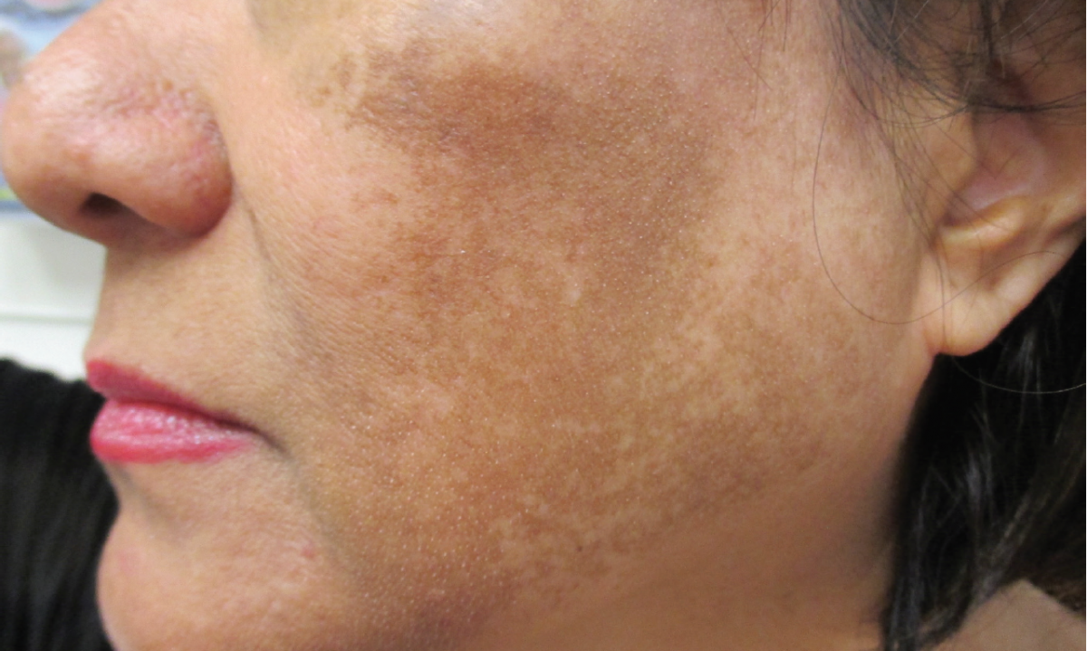
A diagnosis of melasma was made. Wood lamp examination showed accentuation of pigmentation suggestive of epidermal pigmentation. The patient was treated with a series of trichloroacetic acid chemical peels, as well as a topical azelaic acid and a retinoid-hydroquinone-steroid (modified Kligman) formulation with significant, albeit incomplete, improvement.
Melasma is an acquired disorder of hypermelanosis characterized by symmetrically distributed hyperpigmented macules/patches involving sun-exposed areas, primarily on the face.1 The term melasma is derived from the Greek word melas, meaning black. Synonyms include chloasma and “mask of pregnancy” when the condition arises during pregnancy.2 The term chloasma is derived from the Greek word chloazein, meaning green.
Epidemiology
Melasma is rarely reported before puberty and is most common in women of reproductive age, especially in their 30s and 40s.3-5 The condition affects all racial groups but is most prevalent in dark-skinned individuals with Fitzpatrick skin types IV to VI, especially Hispanic/Latino, Asian, and African American persons.3-7 The reported prevalence is 4% to 10% among Latina women in the United States.8,9 The prevalence is as high as 40% in Southeast Asian women.9,10 The female-to-male ratio is approximately 9 to 1.6,11
Etiopathogenesis
The pathogenesis is complex and involves the interplay of various factors, the most important one being chronic UV exposure.3,12,13 Other risk factors include genetic predisposition, pregnancy, thyroid dysfunction, use of certain scented cosmetics, oral contraceptive pills, hormonal therapy, photosensitizing agents, and medications (eg, phenytoin, imatinib, amiodarone, tetracyclines).2,4,7,11,14
UV radiation stimulates melanogenesis by direct effects on melanocytes and by indirect effects on keratinocytes releasing melanogenic factors.10,15 It has been shown that UV radiation induces α-melanocyte-stimulating hormone and melanocortin within melanocytes and keratinocytes which help to up-regulate melanocyte proliferation and melanogenesis.1,2,12,15 Lesional melanocytes contain an increased number of melanosomes compared with melanocytes from adjacent normal-looking skin.7 The resultant increase in the number and activity of melanocytes enhances formation and transfer of melanosomes to epidermis and dermis.8 It has also been shown that fibroblasts from melasma lesions secrete more melanogenic cytokines such as nerve growth factor-β than those in normal skin and thus may play a role in melanogenesis and the pathogenesis of melasma.9,16 Overexpression of cadherin 11 (CDH11), stem cell factor, proto-oncogene receptor tyrosine kinase (KIT), and vascular endothelial growth factor in fibroblasts and keratinocytes could induce basement membrane disruption and dermal changes, independent of UV radiation.1,17
Histopathology
Three histologic patterns of pigmentation have been described: epidermal, dermal, and mixed. In the epidermal type (most common), there is an increase of melanin deposits in the basal and suprabasal epidermal layers.8 In the dermal type, many melanin-laden macrophages are seen in the superficial dermis, often surrounding perivascular spaces.8 The mixed type shows a combination of the 2 patterns. Other histologic findings include basement membrane disruption and dermal changes (solar elastosis, increased number and size of blood vessels, and increased number of mast cells).3,17
Clinical Manifestations
Typically, melasma presents with asymptomatic, symmetrically distributed, well-demarcated macules/patches with serrated, irregular, and geographic borders.5,11 The macules/patches usually develop gradually over time. Pigmentation can be guttate, confetti-like, linear, or confluent.5 The color varies from light brown to dark brown to bluish gray.5 Epidermal melasma is usually light brown, and Wood light enhances the color contrast between hyperpigmented areas and normal skin.18 Dermal melasma tends to be bluish or grayish due to the Tyndall effect and exhibits no accentuation of color contrast under Wood light.18 The mixed type is usually dark brown with variable enhancement on Wood light examination.18
Melasma occurs mainly on sun-exposed areas, mostly on the face, less commonly on the forearms, and rarely on the upper chest.1,10,19 Three clinical patterns of pigment distribution are recognized, namely centrofacial (65%), malar (20%), and mandibular (15%).11,12 The centrofacial pattern involves the forehead, cheeks, upper lip, nose, and chin. The malar pattern involves the cheeks and nose.9 The mandibular pattern involves the ramus of the mandible.9
Clinical and Quality of Life Assessment
The Melasma Area and Severity Index (MASI) can be used to assess the severity of the condition and used as an outcome measure for melasma studies.20,21 The MASI score is calculated by subjective assessment of 3 factors—namely, area of involvement, darkness, and homogeneity—with the forehead, right malar region, left malar region, and chin corresponding to 30%, 30%, 30%, and 10% of the total face, respectively.20 The area of involvement in each of these 4 areas is given a numeric value of 0 to 6 (0, no involvement; 1, < 10%; 2, 10% to 29%; 3, 30% to 49%; 4, 50% to 69%; 5, 70% to 89%; and 6, ≥ 90%). Darkness and homogeneity are rated on a scale from 0 to 4 (0, absent; 1, slight; 2, mild; 3, marked; and 4, maximum). The reliability and validity of MASI was tested by Pandya and colleagues, who recommended using a modified MASI (mMASI) with elimination of the homogeneity component.22
The most commonly used tool that reliably measures the effect of melasma on quality of life is the MelasQol.10,23 The MelasQol is a questionnaire containing 10 questions regarding the impact of melasma on the emotional condition, social relationships, and daily activities of patients.6,23 The patient ranks on a scale of 1 (not bothered at all) to 7 (bothered all the time) how he or she feels about the melasma.23 The total score is calculated by the sum of all scales for each question (total score ranges from 10 to 70). The tool has been validated for different countries and translated into Spanish, Turkish, French, and Portuguese.6
Diagnosis
The diagnosis is a clinical one. Light yellow-brown uniform macules/patches and dark brown macules/patches are 2 major dermoscopic features of melasma. In vivo reflectance confocal microscopy can be used to define the presence and location of pigment in melasma.24 Skin biopsy is infrequently warranted if the diagnosis is in doubt.
Differential Diagnosis
Among the differential diagnoses are postinflammatory hyperpigmentation, drug-induced hyperpigmentation, ephelides (freckles), solar lentigines, lentigo simplex, actinic dyschromia, exogenous ochronosis, nevus of Ota, nevus of Hori, poikiloderma of Civatte, Becker melanosis, café au lait macules/patches, nevus spilus, actinic lichen planus, frictional melanosis, tinea versicolor, linear and whorled nevoid hypermelanosis, and flat seborrheic keratoses.9,18
Complications
Melasma can be esthetically displeasing and socially debilitating. This is particularly true of facial lesions. The condition can have a negative impact on the quality of life, leisure activities, social life, and emotional well-being and may result in psychological disturbances.1,6,8,25
Prognosis
The prognosis depends on the risk factors and the histologic subtype. Melasma may disappear or improve significantly several months postpartum or after cessation of offending agents such as oral contraceptives. While epidermal pigmentation tends to respond better to depigmenting agents, dermal pigmentation responds variably and often unsatisfactorily.5 Recurrence is common, especially with reexposure to sunlight.1
Prevention
Avoidance of sun exposure, regular use of broad-spectrum sunscreens, and wearing of protective wide-brimmed hats and clothing when outdoors should be emphasized.26 Offending agents such as certain scented cosmetics, oral contraceptives, and photosensitizing agents should be avoided if possible.
Treatment
Treatment of melasma involves the use of topical depigmenting agents such as hydroquinone, retinoic acid, ascorbic acid, corticosteroids, kojic acid, glycolic acid, licorice root extract, resorcinol, and azelaic acid.1-4,10 Physical therapies such as chemical peels (eg, glycolic acid, trichloroacetic acid, salicylic acid, lactic acid), dermabrasion, lasers, and intense pulsed light have been used with varying degrees of success.3,4 Unfortunately, the results of monotherapies are usually disappointing, especially in dark-skinned individuals, who are are especially difficult to treat because of the increased risk of postinflammatory hyperpigmentation.10 Combinations of agents or modalities are often used, especially in refractory cases.1 The current limited evidence supports the efficacy and use of a triple combination product (modified Kligman formulation) with topical hydroquinone, a retinoid, and a corticosteroid, which is more effective than treatment with any 1 or 2 of the ingredients.4,26,27 Camouflage make-up is an important component of melasma management.10,26 Recent studies have shown that oral tranexamic acid may be a valuable adjunct in the treatment of refractory melasma.28,29
Alexander K. C. Leung, MD, is clinical professor of pediatrics at the University of Calgary and a pediatric consultant at the Alberta Children’s Hospital in Calgary, Alberta, Canada.
Benjamin Barankin, MD, is a dermatologist and the medical director and founder of the Toronto Dermatology Centre in Toronto, Ontario, Canada.
REFERENCES:
- Goldstein BG, Goldstein AO, Callender VD. Melasma. UpToDate. http://www.uptodate.com/contents/melasma. Updated June 13, 2017. Accessed July 7, 2017.
- Pandya A, Berneburg M, Ortonne J-P, Picardo M. Guidelines for clinical trials in melasma. Br J Dermatol. 2007;156(suppl 1):21-28.
- Kwon S-H, Hwang Y-J, Lee S-K, Park K-C. Heterogeneous pathology of melasma and its clinical implications. Int J Mol Sci. 2016;17(6):824.
- Dunbar S, Posnick D, Bloom B, Elias C, Zito P, Goldberg DJ. Energy-based device treatment of melasma: an update and review of the literature. J Cosmet Laser Ther. 2017;19(1):2-12.
- Ting PT, Barankin B. Can you identify this condition? Melasma. Can Fam Physician. 2005;51:353-355.
- Ikino JK, Nunes DH, Silva VPM, Fröde TS, Sens MM. Melasma and assessment of the quality of life in Brazilian women. An Bras Dermatol. 2015;90(2):196-200.
- Rostami Mogaddam M, Iranparvar Alamdari M, Maleki N, Safavi Ardabili N, Abedkouhi S. Evaluation of autoimmune thyroid disease in melasma. J Cosmet Dermatol. 2015;14(2):167-171.
- Ball Arefiev KL, Hantash BM. Advances in the treatment of melasma: a review of the recent literature. Dermatol Surg. 2012;38(7 pt 1):971-984.
- Sheth VM, Pandya AG. Melasma: a comprehensive update: part I. J Am Acad Dermatol. 2011;65(4):689-697.
- Rodrigues M, Pandya AG. Melasma: clinical diagnosis and management options. Australas J Dermatol. 2015;56(3):151-163.
- Nicolaidou E, Katsambas AD. Pigmentation disorders: hyperpigmentation and hypopigmentation. Clin Dermatol. 2014;32(1):66-72.
- Cestari TF, Dantas LP, Boza JC. Acquired hyperpigmentations. An Bras Dermatol. 2014;89(1):11-25.
- Handel AC, Lima PB, Tonolli VM, Miot LDB, Miot HA. Risk factors for facial melasma in women: a case-control study. Br J Dermatol. 2014;171(3):588-594.
- Ghunawat S, Sarkar R, Garg VK. Imatinib induced melasma-like pigmentation: report of five cases and review of literature. Indian J Dermatol Venereol Leprol. 2016;82(4):409-412.
- Lee A-Y. Recent progress in melasma pathogenesis. Pigment Cell Melanoma Res. 2015;28(6):648-660.
- Byun JW, Park IS, Choi GS, Shin J. Role of fibroblast-derived factors in the pathogenesis of melasma. Clin Exp Dermatol. 2016;41(6):601-609.
- Kim N-H, Choi S-H, Lee TR, Lee C-H, Lee A-Y. Cadherin 11 involved in basement membrane damage and dermal changes in melasma. Acta Derm Venereol. 2016;96(5):635-640.
- Vashi N, Kundu R. Acquired hyperpigmentation disorders. UpToDate. http://www.uptodate.com/contents/acquired-hyperpigmentation-disorders. Updated June 16, 2017. Accessed July 7, 2017.
- Madke B, Kar S, Yadav N, Bonde P. Extrafacial melasma over forearms. Indian Dermatol Online J. 2016;7(4):344-345.
- Kimbrough-Green CK, Griffiths CEM, Finkel LJ, et al. Topical retinoic acid (tretinoin) for melasma in black patients: a vehicle-controlled clinical trial. Arch Dermatol. 1994;130(6):727-733.
- Rodrigues M, Ayala-Cortés AS, Rodríguez-Arámbula A, Hynan LS, Pandya AG. Interpretability of the Modified Melasma Area and Severity Index (mMASI). JAMA Dermatol. 2016;152(9):1051-1052.
- Pandya AG, Hynan LS, Bhore R, et al. Reliability assessment and validation of the Melasma Area and Severity Index (MASI) and a new modified MASI scoring method. J Am Acad Dermatol. 2011;64(1):78-83, 83.e1-83.e2.
- Balkrishnan R, Mcmichael AJ, Camacho FT, et al. Development and validation of a health-related quality of life instrument for women with melasma. Br J Dermatol. 2003;149(3):572-577.
- Costa MC, Eljaiek HV, Abraham LS, Azulay-Abulafia L, Ardigo M. In vivo reflectance confocal microscopy in a typical case of melasma. An Bras Dermatol. 2012;87(5):782-784.
- Harumi O, Goh CL. The effect of melasma on the quality of life in a sample of women living in Singapore. J Clin Aesthet Dermatol. 2016;9(1):21-24.
- Sheth VM, Pandya AG. Melasma: a comprehensive update: part II. J Am Acad Dermatol. 2011;65(4):699-714.
- Jutley GS, Rajaratnam R, Halpern J, Salim A, Emmett C. Systematic review of randomized controlled trials on interventions for melasma: an abridged Cochrane review. J Am Acad Dermatol. 2014;70(2):369-373.
- Lee HC, Thng TGS, Goh CL. Oral tranexamic acid (TA) in the treatment of melasma: a retrospective analysis. J Am Acad Dermatol. 2016;75(2):385-392.
- Tan AWM, Sen P, Chua SH, Goh BK. Oral tranexamic acid lightens refractory melasma [published online May 13, 2016]. Australas J Dermatol. doi:10.1111/ajd.12474