
Peer Reviewed
Assessing Upper Gastrointestinal Bleeding in an 88-Year-Old Man With Hematemesis
Author:
Ronald N. Rubin, MD—Series Editor
Citation:
Rubin RN. Assessing upper gastrointestinal bleeding in an 88-year-old man with hematemesis. Consultant. 2017;57(8):483-484.
An 88-year-old man was awakened by upper abdominal pain and nausea. Before long, he began to experience hematemesis, prompting his wife to call for an ambulance, which transported him to the emergency department (ED). In the ED, he denied syncope or dizziness. He reported having had similar but much milder abdominal pains, along with black bowel movements, in the 2 days leading up to the current event.
The man’s health was very good considering his age. His mild hypertension was well-controlled with metoprolol, and he also took a statin. He had never had a myocardial infarction or congestive heart failure. He rarely drank alcohol and had no history of liver disease. He had been taking a daily adult aspirin for 6 months after reading that it prevents heart attack and stroke, and his family physician did not vigorously oppose it.
On physical examination, he was somewhat clammy and cool. His pulse was 108 beats/min, and his systolic blood pressure was 95 mm Hg (he was not tested for postural change). Oxygen saturation was 97% on room air. There was mild tenderness to direct deep palpation of the epigastrium without rebound. There were no stigmata of chronic liver disease.
Results of a guaiac test showed that his stool was melanotic and strongly heme-positive. Blood tests revealed a hemoglobin (Hb) level of 11.2 g/dL, a creatinine level of 1.3 mg/dL, and a blood urea nitrogen level of 34 mg/dL.
Answer and discussion on next page
Answer: C is the incorrect statement
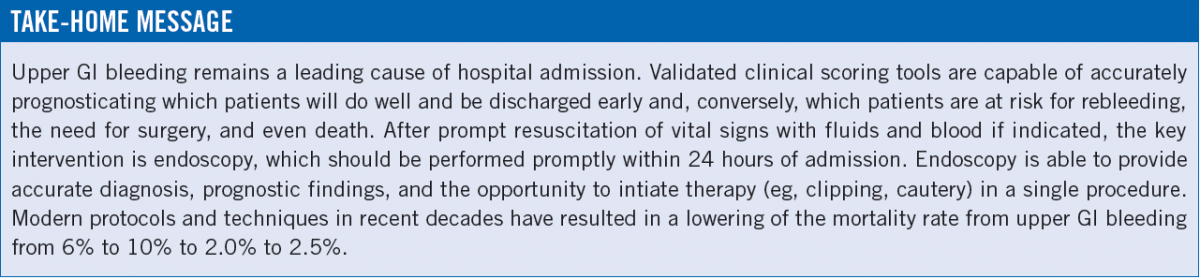
Upper GI bleeding is one of the most dramatic and common clinical presentations of disease and is the most frequent reason for hospital admission related to GI illness. Some good news is that after many decades, the fatality rate of such acute events has finally decreased from the very stubborn rate of 6% to 10% to the far lower 2.1% to 2.5% range.1,2
While many lesions can cause GI bleeding, the most frequent cause is peptic ulcers, which have 2 main etiologies: Helicobacter pylori infection and the use of nonsteroidal anti-inflammatory drugs.2 The case vignette here serves as entry for a discussion along these lines.
The first issue is determining the optimal, evidence-based, early approach to stabilize the patient in this dangerous situation, and thereafter to quickly and accurately diagnose the cause of GI bleeding and develop a treatment strategy. Validated scoring schemes are available that use simple clinical parameters and that quite accurately adjudicate the risk of mortality and other serious sequelae, such as the need for surgery and the risk of rebleeding. The most common is the Glasgow-Blatchford score (GBS),3 which assigns points for blood urea nitrogen level, Hb level, systolic blood pressure, heart rate, and the presence or absence of melena, syncope, hepatic disease, and congestive heart failure. These variables are used to generate a number for the patient at presentation. The higher the number, the higher the probability of a poor outcome—rebleeding, the need for surgery, or death.4 The patient presented here had a GBS of 11, a serious number made more so by his advanced age.
The first goal is stabilization of vital signs. The relatively solid value of the patient’s Hb at admission should not provide false comfort, since patients bleed whole blood, and only hours later, after plasma shifts into the vascular space, is the true Hb deficit and value more clearly obvious. So whereas a baseline very lowered Hb (< 10 g/dL, representing 6 GBS points) is a genuine reason for alarm, early on, other Hb levels do not predict the severity of bleeding; thus, Answer C is not correct.
Regarding if and when to initiate blood transfusion, good trials have shown that a trigger value of 7.0 g/dL is noninferior to a trigger of 9.0 g/dL in hemodynamically stable patients.5 There is much elasticity in clinical practice, however, with many clinicians having a somewhat higher threshold when hemodynamic instability or cardiovascular disease (or both) are present.
The next issue is the use of GI pharmacologic agents (including timing, route, and duration) and endoscopy. Although the PPI use is almost reflex-like, large meta-analyses of trials of early and aggressive PPI therapy did not show a significant reduction in the key outcomes of rebleeding, the need for surgery, or death.1,4,6 This topic is quite argued over in the GI literature, with extreme variability in clinical practice and even among the published guidelines. For now, on a literature analysis basis, Answer A is correct—acute administration of a PPI and erythromycin may help reduce the incidence of various secondary end points but not mortality or the need for surgery.
Evidence for the use of endoscopy is far more clear and uniform. It is the keystone of early management of upper GI bleeding and likely is why the mortality rate has decreased so significantly. Prompt endoscopy within 24 hours of admission (and, very importantly, after adequate resuscitation of vital signs and the Hb level) has been demonstrated in multiple excellent studies to positively impact rebleeding, the need for surgery, and death.1,4 Thus, Answer B also is a correct statement.
Also of great clinical importance, endoscopy definitively diagnoses the precise lesion causing the bleeding and indicates intermediate and even long-term treatment strategies,1,4 making Answer D a correct statement, as well. Classic findings such as the active bleeding, spurting, and visible vessels indicates the need for endoscopic therapies such as clipping, which can be done during the same endoscopy procedure. Biopsies can and should be performed during endoscopy to reveal the presence or absence of malignancy or infection with H pylori, each of which has its own clinical treatment pathway.
Patient Follow-Up
The patient underwent acute resuscitation with several liters of saline. A second complete blood cell count showed that the Hb level had declined to 8.9 g/dL, and he received a transfusion of 2 units packed red blood cells. Results of electrocardiography and cardiac troponin tests were negative for ischemia. After his blood pressure and Hb level stabilized, that morning he underwent urgent endoscopy, which revealed a well-circumscribed, clean-based, 1.5-cm duodenal ulcer and a scattering of gastritis lesions. There was minimal oozing and no visible vessels or adherent clots. Biopsies revealed areas of gastritis, no malignancy, and negative test results for H pylori infection. The patient was treated with an oral PPI for 3 days. He remained clinically stable, as did his Hb level, and he was discharged home on day 3 on a once-daily PPI regimen.
Ronald N. Rubin, MD, is a professor of medicine at the Lewis Katz School of Medicine at Temple University and is chief of clinical hematology in the Department of Medicine at Temple University Hospital in Philadelphia, Pennsylvania.
REFERENCES:
- Laine L. Upper gastrointestinal bleeding due to a peptic ulcer. N Engl J Med. 2016;374(24):2367-2376.
- Laine L, Yang H, Chang S-C, Datto C. Trends for incidence of hospitalization and death due to GI complications in the United States from 2001 to 2009. Am J Gastroenterol. 2012;107(8):1190-1195.
- Laursen SB, Dalton HR, Murray JA, et al; Upper Gastrointestinal Hemorrhage International Consortium. Performance of new thresholds of the Glasgow Blatchford score in managing patients with upper gastrointestinal bleeding. Clin Gastroenterol Hepatol. 2015;13(1):115-121.e2.
- Laine L, Jensen DM. Management of patients with ulcer bleeding. Am J Gastroenterol. 2012;107(3):345-360.
- Villanueva C, Colombo A, Bosch A, et al. Transfusion strategies for acute upper gastrointestinal bleeding. N Engl J Med. 2013;368(1):11-21.
- Barkun AN, Bardou M, Kuipers EJ, et al; International Consensus Upper Gastrointestinal Bleeding Conference Group. International consensus recommendations on the management of patients with nonvariceal upper gastrointestinal bleeding. Ann Intern Med. 2010;152(2):101-113.