
Peer Reviewed
The Approach to an Abnormal Platelet Count
Author:
Ronald N. Rubin, MD—Series Editor
Citation:
Rubin RN. The approach to an abnormal platelet count. Consultant. 2017;57(10):608a,608h.
A 57-year-old woman recently had a complete blood cell count (CBC), the results of which returned with an abnormal platelet count of 575 × 103/µL. The red blood cell (RBC) count, white blood cell (WBC) count, and mean corpuscular volume (MCV) were normal.
Upon review of her medical record, her platelet count had been slightly but consistently elevated (450 to 525 × 103/µL) for at least 2 years. No specific complaints had prompted her visit—it was for a routine examination, blood testing, and a mammogram, all of which had normal or negative results, save for the elevated platelet count. She had not been hospitalized since childbirth 21 years ago. Her past medical and surgical history was noncontributory. Her only medication was the sporadic use of nonsteroidal anti-inflammatory drugs for soreness after activity such as tennis and working out at the gym. There was no family history of blood disorders or bleeding diathesis.
Physical examination findings were all normal, including the absence of petechiae or purpura and splenomegaly. A CBC revealed a hemoglobin level of 13.0 g/dL, an MCV of 86 µm3, a WBC count of 9000/µL with a normal differential count, and a platelet count of 574 × 103/µL. Her ferritin level was normal at 90 ng/mL, and results of a basic metabolic panel were normal.
Answer and discussion on next page
Answer: C is the correct statement
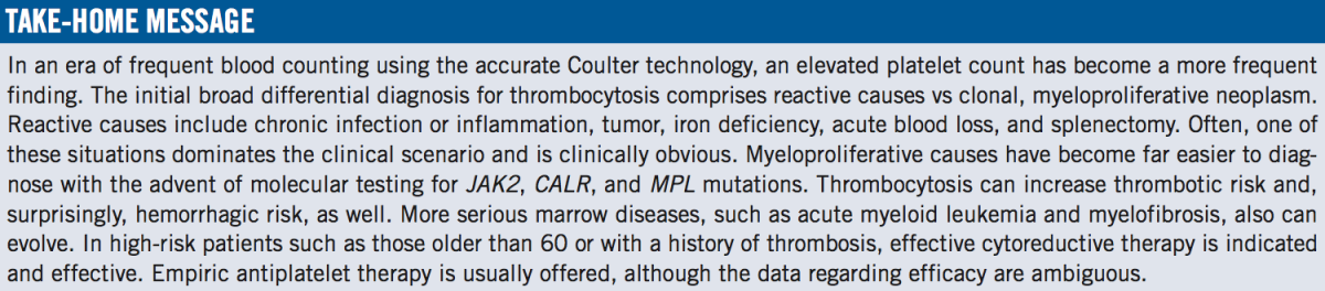
The accurate measurement of platelets and the evaluation of elevated platelet counts have come a very long way in recent years. At one time, technicians estimated the platelet number by viewing a blood slide and then checking a box indicating whether the count was “decreased,” “adequate,” or “increased.” Precise numbers required dilution techniques and technician time that did damage to the laboratory budget. Advances in Coulter counter machine technology have made accurate platelet counts readily and cheaply available. Not surprisingly, thrombocytosis has become far more commonly detected, such that cases like the one presented here are a frequent clinical situation.
The patient here had a platelet count of 574 × 103/µL, well above the normal range of 150 × 103/µL and 400-450 × 103/µL. Other measurements dating back many months indicate that the elevation was a bona fide abnormality.
The initial dividing point in the differential diagnosis is either reactive thrombocytosis (the bone marrow is intrinsically normal and is appropriately responding to a physiologic increased thrombopoietin signal) or autonomous thrombocytosis (the marrow has lost its normal response physiology and is making platelets “on its own”). The latter situation is a reflection of malignancy and can now be identified by demonstrating the presence of monoclonal antibodies as molecular markers in bone marrow cell precursors. Besides demonstrating the presence of a clonal malignancy, however subtle and clinically mild, therapeutics for and management of platelet counts and platelet function differ between reactive and malignant causes.
Reactive thrombocytosis is seen in several well-defined situations. The classic ones include post-splenectomy, serious chronic infections or inflammations such as osteomyelitis and collagen vascular diseases, severe iron deficiency, and acute hemorrhage. Most often, the cause of the thrombocytosis dominates the clinical picture—for example, a surgical intensive-care unit patient with abdominal abscess and multiple surgical procedures, with a worrisome incidental finding of an elevated platelet count flagged during Friday-afternoon rounds. In these reactive situations, platelet count elevation to greater than 1 million/µL (1000 × 103/µL) can occur, such that the number itself does not have value in differentiating reactive disease from clonal disease.
A long history of detailed observational studies has documented that reactive thrombocytosis has no morbidity related to platelet count and requires neither platelet-lowering nor platelet-inhibiting therapies.1 Once the stimulus has been addressed, the platelet count will normalize, usually within several weeks. Our patient had a benign past medical and surgical history and no findings suggestive of any of the usual causes of reactive hrombocytosis. Furthermore, she had a long history of platelet elevations such that an occult neoplasm or infection would have declared by now. Her ferritin level and MCV were within normal limits, excluding iron deficiency as a cause, so Answer B, a trial of intravenous iron therapy, is not the optimal maneuver.
The clonal disorders causing elevated platelet counts (historically referred to as thrombocythemias) are members of the myeloproliferative disease family and include essential thrombocythemia (ET), polycythemia vera (PV), and myelofibrosis. Formerly, the diagnosis was an operational one—a workup to exclude the reactive causes with ferritin level assessment, computed tomography scans, and bone marrow tests; when results are negative, assume myeloproliferative causation. Today we can prospectively and definitively make the diagnosis using genetic markers to demonstrate the clonal presence of abnormal genes. Specifically, these include JAK2 V617F and JAK2 exon 12 mutations (found in 95% of PV cases and 60% of ET cases), mutations of molecular chaperone gene CALR (20%-25% of ET cases), and mutations of thrombopoietin receptor gene MPL (5% of ET cases).2 Molecular testing for these mutations is indicated in our patient, making Answer C the correct choice.
The remaining answers concern the management of myeloproliferative causes of thrombocytosis. Unlike reactive situations, myeloproliferative situations have associated morbidity that may need to be addressed, although the prognosis for these neoplasms is quite good, with survival measured in decades in most cases. The morbidities involve thrombotic and/or hemorrhagic risk due to functional derangements associated with myeloproliferative platelets, and a natural history of degeneration of PV and ET into more serious marrow disorders such as acute myeloid leukemia or myelofibrosis in approximately 5% of cases.2,3 Certain risk factors are associated with increased thrombotic risk, such as age above 60 years, a history of thrombotic disease, and, in some studies, a platelet count above 1 million/µL (although the data relating thrombotic risk to platelet count are the weakest among the 3 factors).4,5 In these instances, the standard of care is cytoreductive therapy, usually with the mild, well-tolerated, and nonleukemogenic agent hydroxyurea. Most clinicians would lower the count to the 600 × 103/µL range and not push toward a count in the normal range. In any event, our patient does not have any of the risk factors, so she should be observed for now, making Answer A incorrect on several counts.
Finally, the issue of antiplatelet therapy needs to be discussed. Reasonable data from good studies suggest a role for low-dose aspirin in reducing thrombotic risk in full-blown PV.6 The data for ET are far less impressive, with a recent meta-analysis failing to demonstrate any difference associated with the use of aspirin and casting doubt on the utility of this maneuver.4,5 Based on these findings, Answer D is not correct, especially at the dosage of 325 mg. Having said this, many clinicians for now do continue to initiate a daily 81-mg aspirin regimen at diagnosis, because it seems so easy, nontoxic, and inexpensive, and because it “may help.” The situation begs for a trial (which would not be difficult to do) to definitively answer this question for this growing population of patients.5
Patient Follow-Up
Molecular testing was performed, the results of which revealed the presence of the JAK2 V617F mutation in exon 14, confirming a myeloproliferative cause of the patient’s elevated platelet count. Since she was younger than 60 and had no history of thrombotic events (and additionally because the platelet count itself was not extremely elevated), no cytoreductive therapy was considered. After a long and detailed discussion of the potential risks and benefits of antiplatelet agents, the patient expressed a strong preference for them, and a daily 81-mg aspirin regimen was started. At 6 months follow-up, she had experienced no adverse events from the aspirin, and her CBC results were essentially unchanged, with a platelet count of 469 × 103/µL.
Ronald N. Rubin, MD, is a professor of medicine at the Lewis Katz School of Medicine at Temple University and is chief of clinical hematology in the Department of Medicine at Temple University Hospital in Philadelphia, Pennsylvania.
References:
- Schafer AI. Thrombocytosis. N Engl J Med. 2004;350(12):1211-1219.
- Spivak JL. Myeloproliferative neoplasms. N Engl J Med. 2017;376(22):2168-2181.
- Tefferi A, Barbui T. Essential thrombocythemia and polycythemia vera: focus on clinical practice. Mayo Clin Proc. 2015;90(9):1283-1293.
- Chu DK, Hills CM, Leong DP, Anand SS, Siegal DM. Benefits and risks of antithrombotic therapy in essential thrombocythemia: a systematic review. Ann Intern Med. 2017;167(3):170-180.
- Bennett JS. Are antiplatelet agents beneficial in essential thrombocythemia? Maybe yes, probably no. Ann Intern Med. 2017;167(3):206-207.
- Landolfi R, Marchioli R, Kuttie J, et al; European Collaboration on Low-Dose Aspirin in Polycythemia Vera Investigators. Efficacy and safety of low-dose aspirin in polycythemia vera. N Engl J Med. 2004;350(2):114-124.