
Peer Reviewed
An Active 70-Year-Old Man With Increasing Fatigue Upon Exertion
Author:
Ronald Rubin, MD—Series Editor
Citation:
Rubin R. An active 70-year-old man with increasing fatigue upon exertion. Consultant. 2016;56(12):1112-1113.
A 70-year-old man presents with increasing fatigue with activity. He is an avid and talented golfer and a senior softball player who has played regularly for many years. In recent months, he has noted profound fatigue in the last holes of a golf round and in the latter innings of a softball game.
Specific questioning reveals probable dyspnea on vigorous exertion, which is different and new from that of prior months. He has had no chest pain, paroxysmal nocturnal dyspnea, or significant pedal edema. He has had mild hypertension that previously had been well controlled with an angiotensin-converting enzyme inhibitor (ACEI) and a diuretic, which had been titrated to a higher dosage 3 weeks ago by his primary physician without any noticeable improvement in symptoms. The man is a nonsmoker and is not diabetic. He did have an episode of paroxysmal atrial fibrillation (AF) 6 years ago that had been treated with electrical cardioversion without recurrence or sequelae since.
Physical examination reveals a well-appearing man with a heart rate of 120 beats/min and a blood pressure of 105/70 mm Hg. The head, eyes, ears, nose, and throat are normal on examination. There are no neck masses, and carotid pulsations are normal. The chest is clear to percussion and auscultation. The heart has a regular rhythm of 120 beats/min with no gross murmurs or gallops. Abdominal examination is negative for masses or organomegaly. The skin has no rashes or petechiae. There is no pedal edema.
Electrocardiography (ECG) reveals atrial flutter with a regular ventricular rate of 120 beats/min. Transthoracic echocardiography (TTE) shows no valvular lesions and no regional wall abnormalities, but it does show a slightly dilated heart and an ejection fraction (EF) of 25%.
Results of continuous 48-hour Holter monitoring of the heart rate revealed essential continuous atrial flutter, with heart rates between 120 and 124 beats/min.
Answer and discussion on next page.
Answer: D, perform TEE, and if negative for atrial thrombus, proceed to early electrical cardioversion.
In addition to the usual and classic symptoms of CHF—dyspnea with exertion and pedal edema—patients with CHF often have an accompanying tachycardia, usually sinus tachycardia. However, on occasion tachyarrhythmia may develop first, and if it is unrecognized or not successfully treated, it can in itself cause the development of cardiac muscle dysfunction over a sustained time and a secondary cardiomyopathy with CHF. By far, the most common arrhythmia in this scenario is AF, but any sustained tachycardia can do this including the less-common atrial flutter, as was the case in this patient. Recognition and appropriate treatment are critical, since the cardiomyopathy related to a sustained tachycardia is reversible in a significant majority of patients if the tachycardia can be controlled.1
The patient’s case described here is a quite typical presentation, which has some unique features compared with many presentations of tachycardia and CHF of other etiology. First, it takes time for the proposed causative derangements of biochemistry and physiology to occur and induce the cardiomyopathy, such that the syndrome is more common with “slower” heart rates in the 120 to 130 beats/min range rather than with more rapid heart rates. The former results in palpitations not being a dominant symptom. Patients with faster heart rates (eg, 150 beats/min) will frequently experience palpitations, which result in presentation and management earlier, before cardiomyopathy can develop.
And, although some history of typical CHF symptomatology can usually be elicited, the presentation of fatigue as a symptom is more common,1 as was seen in the presented case. As stated, heart rates in the 120s are the rule. These can derive from a very wide variety of causative arrhythmias—AF, reentrant supraventricular tachycardia, atrial flutter, and even (although rare) ventricular tachyarrhythmias.1,2
Of course, the ECG is the entry-level diagnostic study and will document the specific arrhythmia and heart rate. Once suspicion of an arrhythmia-based heart problem is in hand, a continuous cardiac monitoring study will confirm the chronicity of the rhythm and the average heart rate being generated. TTE usually is then sufficient to assess overall cardiac function (eg, EF) and exclude other cardiac structural defects with their own specific management (eg, regional wall abnormalities suggesting coronary artery disease or valvular abnormalities). Routine biochemical studies such as immunofixation and iron studies will exclude some of the other more exotic and occult causes of CHF such as amyloidosis and hemochromatosis.
Once the decision is made that the etiology of the cardiomyopathy and CHF is tachycardia-related, appropriate therapy is to definitively reduce the heart rate. The methodology varies and is determined by the specific rhythm and its known behavior. Prompt treatment is important, because the CHF is reversible if this can be done. Standard CHF medical therapy needs to be initiated, continued, or adjusted and includes the proper use of ACEIs, β-blockers, and diuretics. But medical therapy alone for CHF will not be successful, and reversal likely will not occur unless the ventricular rate is addressed, so Answer A does not represent an adequate measure here.
Sometimes, relatively easy maneuvers to slow ventricular rate suffice, particularly in AF, even without restoring normal sinus rhythm.3 For a specifically difficult rhythm, as with our patient’s atrial flutter, the track record of medical antiarrhythmic therapy is poor, and other methods such as electrical cardioversion and ablation come into play. Of the choices offered above, Answer D is preferred to Answer C, given that 2 more months of continued tachycardia would worsen the pathophysiology and adversely affect reversibility, and that normal TEE findings would demonstrate a reduced risk of embolism to a very small and acceptable level.
Finally, the patient should be given the chance to respond to cardioversion prior to undergoing atrioventricular node ablation (Answer B), which requires much further additional collateral therapy (eg, pacemaker placement).2 Thus Answer D seems to be the optimal therapeutic approach at this time in the presented patient’s case.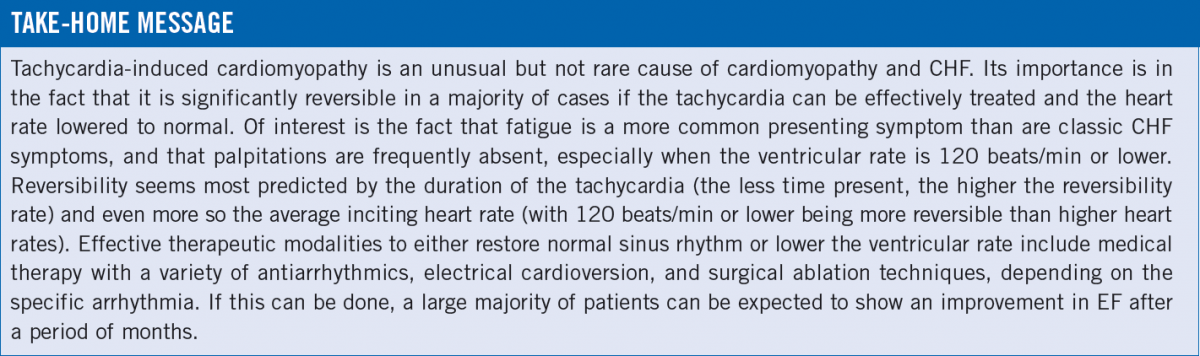
Patient Follow-Up
In the presented patient’s case, results of a complete battery of hematologic and metabolic studies were entirely normal. A TEE confirmed the TTE findings, with the additional data point of no atrial thrombi or atrial appendage thrombi being present. He underwent an elective cardioversion with no complications and thereafter was in sinus rhythm, with a resting heart rate of 76 beats/min. A good regimen of an ACEI, β-blocker, and diuretic was continued.
The patient believes he has had improvement in symptoms at a 4-month follow-up visit; a recent ECG revealed sustained normal sinus rhythm, and a recent echocardiogram showed an EF of 40%. He is back on the golf course and ball field without symptoms.
Ronald Rubin, MD, is a professor of medicine at the Temple University School of Medicine and is chief of clinical hematology in the Department of Medicine at Temple University Hospital in Philadelphia, Pennsylvania.
References:
- Gillette PC, Smith RT, Garson A Jr, et al. Chronic supraventricular tachycardia: a curable cause of congestive cardiomyopathy. JAMA. 1985;253(3):391-392.
- Pizzale S, Lemery R, Green MS, Gollob MH, Tang AS, Birnie DH. Frequency and predictors of tachycardia-induced cardiomyopathy in patients with persistent atrial flutter. Can J Cardiol. 2009;25(8):469-472.
- Roy D, Talajic M, Nattel S, et al; Atrial Fibrillation and Congestive Heart Failure Investigators. Rhythm control versus rate control for atrial fibrillation and heart failure. N Engl J Med. 2008;358(25):2667-2677.