
Peer Reviewed
An 18-Year-Old With Right Knee Pain
AUTHOR:
William Yaakob, MD--Series Editor
CITATION:
Yaakob W. An 18-year-old with right knee pain. Consultant. 2013;55(1):43-44.
An 18-year-old man presents to your office with right knee pain. He says that he has had persistent knee pain for the past 3 weeks since he started playing soccer with his friends in an attempt to lose weight. He said that the knee swells at times, and occasionally, it feels like his knee is catching or locking.
Physical Examination
The patient is a mildly obese young man in no acute distress. Vital signs are within normal limits.
Passive range of motion of the right knee is mildly restricted in knee extension. There is a small joint effusion on the right. You order 2 views of the right knee (Figures 1 and 2).
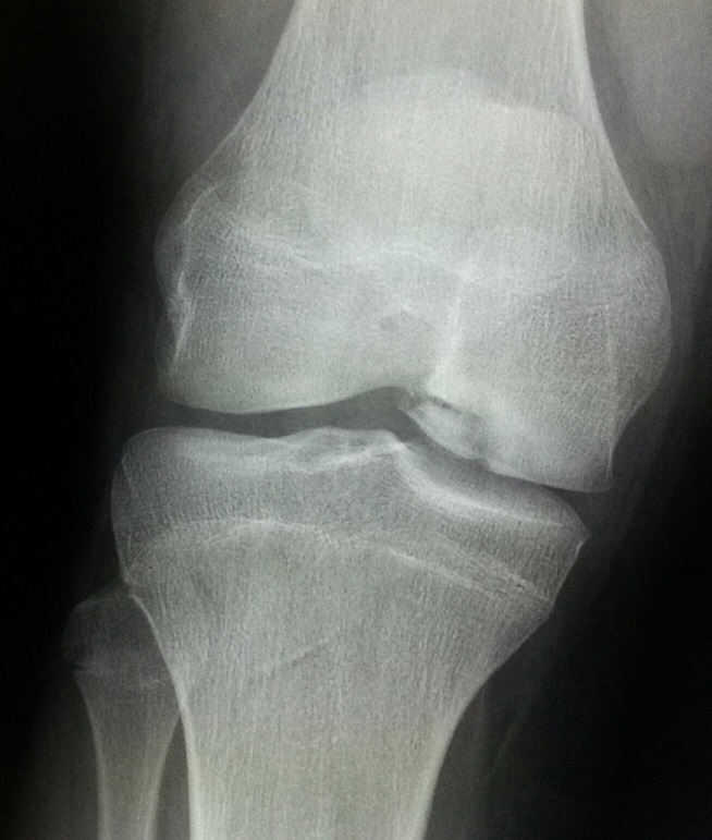
Figure 1. Frontal radiograph of the right knee.
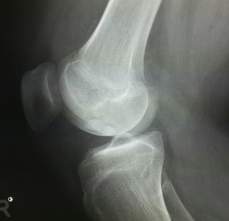
Figure 2. Lateral radiograph of the right knee.
(Answer and discussion on next page)
Answer: Osteochondrosis dessicans
Frontal and lateral radiographs of the right knee demonstrate a region of irregularity of the lateral aspect of the medial femoral condyle at the weight-bearing surface. There is lucency
between this osseous irregularity and the parent bone. On the lateral view, there is a joint effusion. This is the classic appearance and location for osteochondrosis dessicans.
The frontal radiograph of the knee demonstrates cortical irregularity of the lateral aspect of the medial femoral condyle. Upon closer inspection of the region, there is sclerotic region (of increased density) with a band of lucency about it. Increased sclerosis is identified in the adjacent medial femoral condyle. The abnormal region of bone is also small in size compared to what one would expect.
On the lateral radiograph, there is a joint effusion. The abnormality in the medial femoral condyle is visualized (the medial femoral condyle is the larger of the 2 on the lateral view). Again, there is sclerosis of the abnormality with shrinking of the bone in the region of the abnormality. This strongly implies that there is an injury of the overlying cartilage.
Upon further inspection, an MRI was ordered. T1 (Figure 3) and T2 (Figure 4) weighted images were obtained. On the T2 weighted images, there is edema in the adjacent abnormal medial femoral condyle and in the region of sclerosis. The fragment appears to be more displaced laterally on the T1 weighted images versus the T2 weighted images, which strongly implies a loose type of osteochondral injury.
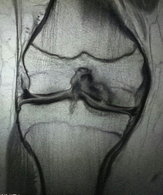
Figure 3. T1 weighted MRI of the right knee in the coronal plane.
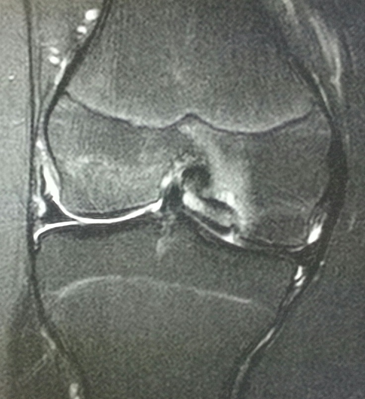
Figure 4. T2 weighted MRI of the right knee in the coronal plane.
Discussion
Osteochondrosis dessicans (OCD) is a type of avascular necrosis in which the blood supply to the bone in the affected area is disrupted with subsequent resorption of bone and fragmentation of the overlying cartilage. The bone deprived of its blood supply shrinks in size and can separate or fragment from the parent bone. The fragments of overlying cartilage can become loose bodies within the joint leading to “catching or locking.”
OCD is of unknown etiology and is seen infrequently. There are approximately 15 to 30 cases per 100,000 seen each year. This is more common in males to females by a ratio of 2-3:1. Although patients are between 10 and 20 years, this can occur at any age. The most common location of involvement is the medial femoral condyle of the knee as shown in this case. OCD can be bilateral in up to 40% of cases.
Outcome of the Case
This patient was referred to orthopedics for treatment. Given that the injury is unstable, surgical fixation of the abnormal fragment from the lateral aspect of the medial femoral condyle to the parent bone is being strongly considered.
William Yaakob, MD, is a board-certified radiologist working in Tallahassee, FL.