Zoon Balanitis in a Middle-aged Man
Zoon balanitis, or plasma cell balanitis, is a chronic, benign, rare inflammatory disorder that manifests as lesions localized on the glans penis and prepuce. The cause is unknown. It typically occurs in middle-aged and elderly uncircumcised men. Here we present the case of a middle-aged man with Zoon balanitis who was treated with topical corticosteroids and tacrolimus.
 THE CASE
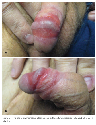
A 54-year-old uncircumcised man presented with erythematous, painful lesions of about 1 year’s duration on the prepuce and glans penis (Figure 1). He had a history of non-Hodgkin lymphoma, type 2 diabetes mellitus, hepatitis C, and hypothyroidism.
The patient was initially treated with clotrimazole 1% cream as well as fluconazole in a single 150-mg dose. No improvement was seen. Bacterial culture and tests for Chlamydia infection and gonorrhea were negative.
A biopsy sample was obtained, which showed changes consistent with Zoon balanitis (Figure 2). The patient was then treated with hydrocortisone valerate 0.2% cream, and the balanitis improved significantly. One month later, the patient was given 0.1% tacrolimus ointment for maintenance therapy. Because he was noncompliant with therapy, the balanitis flared up again; the patient was then given desoximetasone 0.05% ointment.
DISCUSSION
Clinical features. Zoon balanitis, or plasma cell balanitis, was first described in 1952 by Zoon.1 This chronic, benign, rare inflammatory disorder most frequently occurs as lesions localized on the glans penis and prepuce, typically in middle-aged and elderly uncircumcised men.2 These lesions are solitary, sharply demarcated, erythematous, shiny plaques that often have a “cayenne pepper”–speckled appearance. Zoon balanitis is usually asymptomatic but may be associated with pruritus and tenderness.
The cause is unknown; however, it may result from mild trauma, irritation, or friction of the foreskin in a moist environment.3,4 Chronic Mycobacterium smegmatis infection and hypospadias have also been proposed as predisposing factors.5
Differential diagnosis. Zoon balanitis can at times be difficult to distinguish from other erythematous lesions involving the balanopreputial sac (eg, erythroplasia of Queyrat, localized forms of lichen planus, cicatricial pemphigoid, unilesional forms of psoriasis, syphilis, candidiasis), and therefore can be easily misdiagnosed. Other conditions in the differential diagnosis are contact dermatitis, squamous cell carcinoma, lichen sclerosus et atrophicus, and balanitis xerotica obliterans.
Biopsy is necessary to help clarify the nature of the disease. Microscopy for fungi; Tzanck smear; viral, bacterial, and fungal cultures; fasting blood glucose measurement; and serology for syphilis are other tests that may be used to help rule out other causes.
 Histopathology. The histopathological findings of Zoon balanitis are progressive and vary depending on the length of time the disease has been present. Initially, the epidermis can be slightly thickened with patchy lichenoid infiltrate of lymphocytes, parakeratosis, and few plasma cells. As Zoon balanitis progresses, the infiltrate becomes a denser band of multiple plasma-type cells, extravasated erythrocytes, and few neutrophils in the upper epidermis, while the epidermis displays signs of atrophy and superficial erosions. In time, the histology can progress further to show subepidermal clefts, with possible epidermal loss, superficial dermis fibrosis, and siderophage infiltrate.3,5 Diamond-shaped keratinocytes and a uniform spongiosis may be seen.
Treatment. The initial treatment regimen may include good hygiene, emollient creams, and topical corticosteroids. Circumcision is usually curative. Tacrolimus is an alternative treatment for patients in whom there is good reason to avoid topical corticosteroids and for those who refuse surgery.6 Other treatments include carbon dioxide laser,7 erbium:YAG laser,8 copper vapor laser,5 imiquimod,9 fusidic acid,10 and tannic acid.4
1. Zoon JJ. Balanoposthite chronique circonscrite bénigne á plasmcytes. Dermatologica. 1952;105:1-7.
2. Braun-Falco O, Plewig G, Wolff HH, Burgdorf WH, eds. Dermatology. 2nd ed. Berlin: Springer; 2002.
3. Weyers W, Ende Y, Schalla W, Diaz-Cascajo C. Balanitis of Zoon: a clinicopathologic study of 45 cases. Am J Dermatopathol. 2002;24:459-467.
4. Moreno-Arias GA, Camps-Fresneda A, Llaberia C, Palou-Almerich J. Plasma cell balanitis treated with tacrolimus 0.1%. Brit J Dermatol. 2005;153:1204-1206.
5. Santos-Juanes J, Sanchez del Río J, Galache C, Soto J. Topical tacrolimus: an effective therapy for Zoon balanitis. Arch Dermatol. 2004;140:1538-1539.
6. Hernandez-Machin B, Hernando LB, Marrero OB, Hernandez B. Plasma cell balanitis of Zoon treated successfully with topical tacrolimus. Clin Exp Dermatol. 2005;30:588-589.
7. Baldwin HE, Geronemus RG. The treatment of Zoon’s balanitis with the carbon dioxide laser. Dermatol Surg Oncol. 1989;15:491-494.
8. Albertini JG, Holck DE, Farley MF. Zoon’s balanitis treated with erbium:YAG laser ablation. Laser Surg Med. 2002;30:123-126.
9. Nasca MR, De Pasquale R, Micali G. Treatment of Zoon’s balanitis with imiquimod 5% cream. J Drugs Dermatol. 2007;6:532-534.
10. Petersen CS, Thomsen K. Fusidic acid cream in the treatment of plasma cell balanitis. J Am Acad Dermatol. 1992;27(4):633-634.


