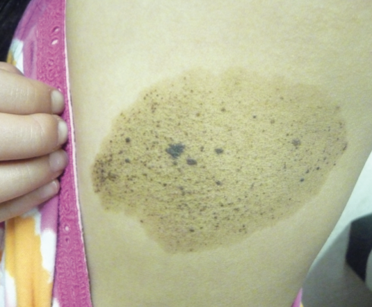
Teenage Girl With Giant Hyperpigmented Patch

HISTORY
A 17-year-old girl presents with an asymptomatic giant hyperpigmented patch on her left upper back. The lesion was first noted when the patient was 6 months old, and it has gradually increased in size over time. Her past medical history is unremarkable. No similar lesion has been observed in other family members.
PHYSICAL EXAMINATION
On examination, there is a sharply circumscribed, light brown hyperpigmented patch on the left upper back. Numerous speckled, dark-brown, hyperpigmented spots are present within the patch. The rest of the examination is unremarkable.
WHAT’S YOURDIAGNOSIS?
(Answer on Next Page)
Answer: Spilus Nevus
Spilus nevus was diagnosed. While the patient was reassured that the lesion is generally benign, follow-up was arranged to monitor for any changes that may suggest potential malignancy.
SPILUS NEVUS: AN OVERVIEW
Spilus nevus, also known as speckled lentiginous nevus, typically presents as a light brown, circumscribed pigmentation that is stippled with dark brown punctate macules or papules.1-5 The word “spilus” is derived from the Greek word spilos, meaning spot.
Spilus nevus occurs in fewer than 0.2% of all newborns, 1.3% to 2.1% of school-aged children, and about 2.3% of adults.6-9 Both sexes are affected equally.7,8,10 There is no racial predominance.8
A spilus nevus may represent a localized defect in neural crest melanoblasts that is influenced by genetic and environmental factors.2,3,7
HISTOPATHOLOGY
Histologic examination reveals an increase in melanin deposition in keratinocytes and an increase in the number of non-nested melanocytes along the basal cell layer of
the epidermis.2,7 Some melanosome macroglobules are found in the background macule and represent melanin autosomes formed through an autophagic process following aberrant melano-genesisis.7 Melanosome macroglobules are characteristic findings in nevus spilus.7 Darker flat speckles represent areas of melanocytic hyperplasia.7 Darker lesions represent nevus cells in a junctional, and/or dermal location.1 Papules consist of melanocytic nests in the papillar and reticular dermis.2,3
CLINICAL MANIFESTATIONS
Although spilus nevus can be congenital, most lesions develop in the first year of life, and some during childhood or adolescence.2,3,7 The lesion often starts as an evenly pigmented light brown macule with few or no speckles. The speckles may appear or increase during childhood or even adulthood.1-3 In the macular type of spilus nevus, the speckles are evenly distributed within the background macule.2 In contrast, the papular variant shows a more scattered and uneven distribution of the speckles within the background macule.2 The diameter of the nevus can range from 1 cm to more than 20 cm.11 In most cases, the lesion is small and solitary.1,12 Sites of predilection include the abdomen and back.3,11 The darker speckled lesions typically number 8 to 10 and are usually 1 to 3 mm in diameter.7
Spilus nevus is usually an isolated finding. Sometimes, however, it may occur as part of phacomatosis spilorosea, phacomatosis pigmentovascularis, phacomatosis pigmentokeratosis, or spilus nevus syndrome.2,3,13
DIFFERENTIAL DIAGNOSIS
Spilus nevus should be differentiated from segmental lentiginosis, Becker’s nevus, linear and whorled nevoid hypermelanosis, and the café au lait spot. In segmental lentiginosis, the background pigmentation is absent.2,3 Becker’s nevus consists of a sharply demarcated, irregular area of hyperpigmentation without any speckled pattern, although hypertrichosis within the lesion is commonly present.2,3 Linear and whorled nevoid hypermelanosis is characterized by linear streaks and swirls of macular hyperpigmentation in a reticulate pattern along the lines of Blaschko without any preceding inflammatory or palpable verruciform lesions. A café au lait spot presents as an ovoid, uniformly brown macule with no overlying darker macules or papules. Café au lait spots are found in 3% to 10% of healthy children and almost all children with neurofibromatosis.14 Occasionally, a café au lait spot in an individual with segmental neurofibromatosis may simulate a spilus nevus.
COMPLICATIONS
Patients with spilus nevi are at low risk for melanoma.7,8,12 Atypical melanocytic proliferations can lead to melanoma formation.1 This is especially true for dysplastic giant congenital nevi and those with an atypical appearance.1 Most reported cases are thin melanomas.7
MANAGEMENT
Although spilus nevi are generally benign, they must be monitored closely so that any changes that suggest malignancy can be detected early.1-3,6,12 This is especially true for dysplastic giant congenital nevi and those with an atypical appearance.9 Dermoscopy and image storage can be helpful for monitoring.12 If there is any doubt, biopsy of the lesion for histological examination should be considered. In most cases, routine prophylactic excision of the lesion is unwarranted. However, prophylactic full-thickness, complete excision of the lesion should be considered in the setting of dysplasia or if an atypical appearance is noted.7
1. Leung AK, Kao CP, Robson WL. A giant congenital nevus spilus in an 8-year-old girl. Adv Ther. 2006;23(5):701-704.
2. Leung AK. Spilus nevus. In: Lang F, ed. The Encyclopedia of Molecular Mechanisms of Disease. Berlin: Springer-Verlag; 2009:1959-1960.
3. Leung AK. Nevus spilus. In: Leung AK, ed. Common Problems in Ambulatory Pediatrics: Specific Clinical Problems. Vol 2. New York: Nova Science Publishers, Inc.; 2011:205-208.
4. Ito M, Hamada Y. Nevus spilus en nappe. Tohuku I Exp Med. 1952;55:44-48.
5. Stewart DM, Altman J, Mehregan AH. Speckled lentiginous nevus. Arch Dermatol. 1978;114(6):895-896.
6. Kaur T, Kanwar AJ. Giant nevus spilus and centrofacial lentiginosis. Pediatr Dermatol. 2004;21(4):516-517.
7. Meguerditchian ANM, Cheney RT, Kane JM III. Nevus spilus with synchronous melanomas: case report and literature review. J Cut Med Surg. 2009;13(2):96-101.
8. Vaidya DC, Schwartz RA, Janniger CK. Nevus spilus. Cutis. 2007;80(6):465-468.
9. Yoneyama K, Kamada N, Mizoguchi M, et al. Malignant melanoma and acquired dermal melanocytosis on congenital nevus spilus. J Dermatol. 2005;32(6):
454-458.
10. Singh S, Jain N, Khanna N, et al. Hairy nevus spilus: a case report. Pediatr Dermatol. 2012;Feb 3: DOI: 10.1111/j.1525-1470.2011.01688.x [Epub ahead of print]
11. Zeren-Bilgin I, Gür S, Aydin O, et al. Melanoma arising in a hairy nevus. Int J Dermatol. 2006;45(11):1362-1364.
12. Corradin MT, Zattra E, Fiorentino R, et al. Nevus spilus and melanoma: case report and review of the literature. J Cut Med Surg. 2010;14(2):85-89.
13. Marti N, Jorda E, Martinez E, et al. Widespread nevus spilus associated with torsion dystonia. Pediatr Dermatol. 2010;27(6):654-656.
14. Leung AK. Café au lait macules. In: Leung AK, ed. Common Problems in Ambulatory Pediatrics: Specific Clinical Problems. Vol 2. New York: Nova Science Publishers, Inc.; 2011:223-227.


