
Peer Reviewed
Mycoplasma pneumoniae–Induced Rash and Mucositis: 2 Pediatric Cases
Authors:
Margaret Rush, MD
Division of Hospitalist Medicine, Children’s National Medical Center, Washington, DCMorgan Leighton, MD
Division of Emergency Medicine, Children’s National Medical Center, Washington, DCAnna Yasmine Kikorian, MD
Division of Dermatology, Children’s National Medical Center, Washington, DCKavita Parikh, MD, MSHS
Division of Hospitalist Medicine, Children’s National Medical Center, Washington, DCCitation:
Rush M, Leighton M, Kikorian AY, Parikh K. Mycoplasma pneumoniae–induced rash and mucositis: 2 pediatric cases. Consultant. 2019;59(6):168-171.Mycoplasma pneumoniae–induced rash and mucositis (MIRM) is a newly categorized clinical entity consisting of prominent mucositis and variable skin involvement in patients with a recent mycoplasma infection. This illness is most prevalent in school-aged boys and may be encountered in outpatient and inpatient settings alike. Although hospitalization may be required for management of dehydration, malnutrition, and pain, the overall morbidity and mortality associated with MIRM is low. We present 2 cases of pediatric patients who were recently hospitalized for MIRM and review the literature regarding presentation, pathophysiology, and management of this illness. It is important to be aware of this new diagnosis in order to recognize MIRM, provide supportive care, consider treatment, and effectively counsel patients and families.
CASE 1
A 16-year-old boy presented with severe mucositis with bullous lesions involving the oral mucosa, lips, and pharynx. He was experiencing an inability to swallow secretions due to severe burning throat pain. He had been also exhibited a low-grade fever with rhinorrhea and a sore throat for the past 3 days. Of note, he had had a previous admission for severe mucositis 3 years prior to presentation, for which he had undergone extensive workup without a clear etiology having been identified. His condition ultimately had improved with supportive care.
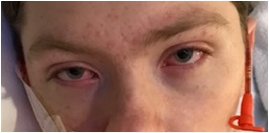
At the current presentation, the patient was admitted to the general pediatric service for treatment and evaluation of mucositis of the conjunctivae, oral mucosa, nares, and urethra, as well as a vesicular skin eruption distributed sparsely over his chest, back, and scrotum (Figures 1 and 2).

Figure 1. Skin findings were notable for sparse vesicular lesions and some atypical target lesions with central vesiculation.
Figure 2. Ocular findings of erythematous conjunctivae due to mucous membrane involvement.A consultant dermatologist suspected MIRM due to the sparsity of the rash and the prodromal viral symptoms. M pneumoniae polymerase chain reaction (PCR) results were positive, and the diagnosis of MIRM was made.
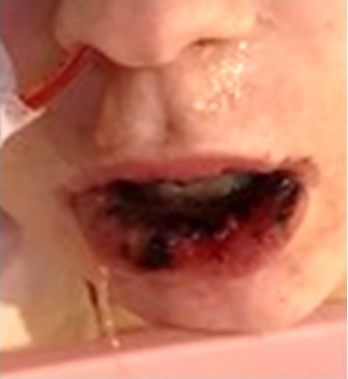
The mucosal lesions progressed during his admission from bullae to erythematous ulcerations with crusting (Figure 3).
Figure 3. Progression of oral mucositis from the first 24 hours of symptoms (top), to approximately 4 days of symptoms (middle), to approximately 1 week of symptoms (bottom).Due to intolerance of feeding and a previous similar episode of unclear etiology, a gastroenterologist was consulted, and the patient underwent upper endoscopy, which showed multiple ulcerated lesions in the esophagus. The patient was hospitalized for 3 weeks with severe mucositis requiring peripheral parenteral nutrition and intravenous pain management. He received a 5-day course of azithromycin and a dose of intravenous immunoglobulin G (IVIG), after which his mucositis and skin lesions resolved, and he was discharged home.
NEXT: Case 2


- Waites KB, Talkington DF. Mycoplasma pneumoniae and its role as a human pathogen. Clin Microbiol Rev. 2004;17(4):697-728.
- Tay Y-K, Huff JC, Weston WL. Mycoplasma pneumoniae infection is associated with Stevens-Johnson syndrome, not erythema multiforme (von Hebra). J Am Acad Dermatol. 1996;35(5 pt 1):757-760.
- Prindaville B, Newell BD, Nopper AJ, Horii KA. Mycoplasma pneumoniae–associated mucocutaneous disease in children: dilemmas in classification. Pediatr Dermatol. 2014;31(6):670-675.
- Bastuji-Garin S, Rzany B, Stern RS, Shear NH, Naldi L, Roujeau J-C. Clinical classification of cases of toxic epidermal necrolysis, Stevens-Johnson syndrome, and erythema multiforme. Arch Dermatol. 1993;129(1):92-96.
- Canavan TN, Mathes EF, Frieden I, Shinkai K. Mycoplasma pneumoniae–induced rash and mucositis as a syndrome distinct from Stevens-Johnson syndrome and erythema multiforme: a systematic review. J Am Acad Dermatol. 2015;72(2):239-24e4.
- Bowling M, Schmutzler T, Glick S. Mycoplasma pneumoniae-induced mucositis without rash in an 11-year-old boy. Clin Case Rep. 2018;6(3):551-552.
- Norton SA. Diagnosing Mycoplasma pneumoniae-induced rash and mucositis (MIRM) in the emergency room. J Am Acad Dermatol. 2015;73(2):e6
- Varghese C, Sharain K, Skalski J, Ramar K. Mycoplasma pneumonia-associated mucositis. BMJ Case Rep. 2014;2014. doi:10.1136/bcr-2014-203795.
- Atkinson TP, Waites KB. Mycoplasma pneumoniae infections in childhood. Pediatr Infect Dis J. 2014;33(1):92-94.
- Martínez-Pérez M, Imbernón-Moya A, Lobato-Berezo A, Churruca-Grijelmo M. Mycoplasma pneumoniae-induced mucocutaneous rash: a new syndrome distinct from erythema multiforme? Report of a new case and review of the literature. Actas Dermosifiliogr. 2016;107(7):e47-e51.
- Santos RP, Silva M, Vieira AP, Brito C. Mycoplasma pneumoniae-induced rash and mucositis: a recently described entity. BMJ Case Rep. 2017;2017. doi:10.1136/bcr-2017-220768.
- Demitsu T, Kawase M, Nagashima K, et al. Mycoplasma pneumoniae-associated mucositis with severe blistering stomatitis and pneumonia successfully treated with azithromycin and infusion therapy. J Dermatol. 2019;46(1):e38-e39.
- Figueira-Coelho J, Lourenço S, Pires AC, Mendonça P, Malhado JA. Mycoplasma pneumoniae-associated mucositis with minimal skin manifestations. Am J Clin Dermatol. 2008;9(6):399-403.
- St. John J, Ratushny V, Liu KJ, et al. Successful use of cyclosporin A for Stevens-Johnson syndrome and toxic epidermal necrolysis in three children. Pediatr Dermatol. 2017;34(5):540-546.
- Ahluwalia J, Wan J, Lee DH, Treat J, Yan AC. Mycoplasma-associated Stevens-Johnson syndrome in children: retrospective review of patients managed with or without intravenous immunoglobulin, systemic corticosteroids, or a combination of therapies. Pediatr Dermatol. 2014;31(6):664-669.
- Ravin KA, Rappaport LD, Zuckerbraun NS, Wadowsky RM, Wald ER, Michaels MM. Mycoplasma pneumoniae and atypical Stevens-Johnson syndrome: a case series. Pediatrics. 2007;119(4):e1002-e1005.