
Peer Reviewed
A 48-Year-Old Man With Fever and Malaise After an International Business Trip
Author:
Ronald N. Rubin, MD—Series Editor
Citation:
Rubin RN. A 48-year-old man with fever and malaise after an international business trip. Consultant. 2018;58(8):221-222.
A 48-year-old man who had had fever and malaise for several days presented for clinical evaluation. He had been well until approximately 3 days previously, when he had awakened at night feeling feverish and in a sweat. These symptoms had been recurring at intervals since then, with a measured temperature of 38.9°C, at times accompanied by rigors and chills. There had been no localizing symptoms such as cough, sputum production, dyspnea, abdominal pains, vomiting or diarrhea, dysuria, or any skin lesions or rash.
The man is an oil company executive who travels frequently and extensively. His most recent trip had been to Nigeria; he had remained in the capital city, Lagos, for most of the time but had ventured into the countryside with his hosts on several occasions. He had been very strict about drinking only bottled water and eating meals at his hotel, with the exception of when he had been a guest in his hosts’ home. He had received all appropriate vaccines prior to his trip, and he had been prescribed malaria prophylaxis. He had returned from Nigeria 28 days ago.
Physical examination revealed a temperature of 37.8°C, a pulse of 106 beats/min, blood pressure of 110/74 mm Hg, and oxygen saturation of 98%. Examination of the head, eyes, ears, nose, and throat was negative for rash or icterus. There were no enlarged lymph nodes in the neck or the supraclavicular or axillary regions. His chest was clear to percussion and auscultation. The heart had regular rhythm and no gallops. There was no abdominal pain or masses. The skin showed no rashes.
Laboratory test results included a normal metabolic panel, save for slight hyponatremia with a sodium level of 132 mEq/L. A complete blood cell count revealed a hemoglobin level of 11.7 g/dL with normal white blood cell and platelet counts. The serum lactate dehydrogenase level was 401 U/L. Chest radiographs were negative for infiltrates.
Find Out!
Answer: A, Falciparum malaria
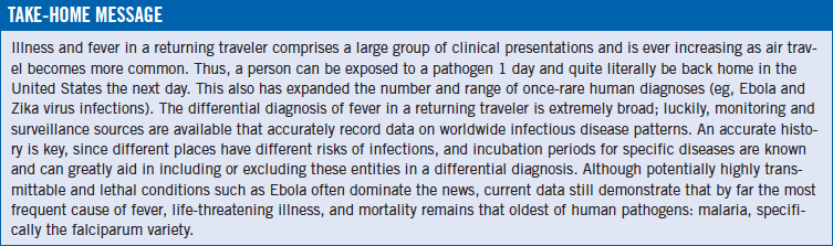
The case presented here exemplifies the ever-more prevalent problem of managing a febrile illness in someone with a recent history of “exotic” travel. The world continues to shrink such that a person can be in a distant tropical area one day and back in Manhattan the next. When such a situation arises, 3 major questions must be answered very quickly. First, is the illness acutely life-threatening to the patient based on an initial risk assessment? Second, and a corollary to the first, what are the transmission risk possibilities to health care personnel and the general population? And third, what is the highest differential diagnosis and, thereafter, the specific diagnosis of the case?
Several excellent tools can be used to gauge where the patient fits into the “febrile traveler” spectrum. Regarding the initial risk assessment, the use of the very easy quick sequential organ failure assessment (qSOFA) score is helpful. This scheme attaches 1 point each for altered mentation, tachypnea, and hypotension. A score of greater than 2 points indicates the need for urgent hospital admission, most likely to an intensive care unit.1
Simultaneously with the initial evaluation is an assessment of the risk of transmission. This is greatly aided by comparing the patient’s travels with information at the Centers for Disease Control and Prevention (CDC) website that monitors global epidemiology and geographic risk data (especially malaria), and at the GeoSentinel surveillance tool developed collaboratively by the CDC and the International Society of Travel Medicine.2,3
Then, once the thorough travel history (eg, places visited, incubation period from visit to fever/symptoms, vaccinations, malarial prophylaxis or other antibiotics), the initial risk assessment, and demographic/epidemiologic data have been obtained, statistics provide further strength. For example, based on recent GeoSentinel data from 1998 to 2011, in patients with acute and life-threatening cases of whom 91% had fever as a dominant symptom, falciparum malaria accounted for 77% of cases, and enteric fevers for 18%; mortality ranged from 0.2% to 0.5%, with malaria being by far the most frequent cause of death.3,4 On this basis alone, Answer A is the correct answer.
Honing down to more-specific data in our case, he had traveled to Nigeria, which is an endemic region for malaria; his incubation period can be calculated to at least 28 days; and he had no gastrointestinal (GI) tract symptoms. His incubation period essentially excludes shorter-incubation diseases such as dengue and Ebola, and the absence of GI symptoms and exposure to unsafe water are strong negatives for enteric fevers.
Dengue is a common infection in tropical areas and is spreading its range to include parts of the United States. It is worldwide in distribution—South Asia, Southeast Asia, South America, Central America, the Caribbean, Africa—anywhere its vector, the Aedes genus of mosquitoes, is found. However, the presented patient lacked the typical symptoms of severe muscle and bone pains (“break-bone fever”) and the commonly seen rash. Most importantly, however, the incubation period of at least 25 days seen in the presented case essentially eliminates dengue as the cause. Thus Answer B is incorrect.
A similar logic essentially excludes Ebola virus (Answer D), as well. Analysis of recent Ebola outbreaks suggests that the illness manifests within 22 days of exposure.1,5 Additionally, the lack of any history of Ebola outbreak in the regions our patient visited and the absence physical contact with infected persons are strong negatives for that diagnosis. Because a diagnosis of Ebola is so significant—life-threatening for the patient, with serious and dangerous public health consequences—it can be argued that testing for Ebola and precautions against its spread are appropriate. However, given the data presented and available, Ebola is not the most likely diagnosis here.
Enteric fever caused by infection with Salmonella enterica subsp enterica ser Typhi and S enterica subsp enterica ser Paratyphi is a possibility that is not excluded by the long incubation period. The illness is transmitted in contaminated food or water by oral transmission cycle and can have an incubation period of 6 to 30 days. Usually, some degree of GI tract symptoms occur along the way, which was not seen in our patient’s case. Enteric fevers account for 18% of severe and life-threatening cases of febrile illness in travelers.3 Current worldwide monitoring data show that almost all cases are contracted in South Asia and Southeast Asia (ie, the Indian subcontinent).2 Our patient’s lack of travel to that area is a strong negative in this case. And despite the vagaries in any patient’s self-reported history, he had no history of having ingesting potentially contaminated food or water “in the bush.” This preponderance of factors makes enteric fever (Answer C) a much less likely etiology here.
NEXT: Patient Follow-Up
Patient Follow-Up
The patient did not manifest any clinical findings of severe disease (qSOFA score, 0). However, there was enough concern for the possible presence of a life-threatening tropical infection to prompt hospital admission. He was placed in isolation in a single room. The first tests performed were thick and thin blood films, which were examined by the hematology and pathology services. The smears demonstrated the presence of Plasmodium falciparum, as did the results of a rapid antigen detection test.
The patient received an artemisinin-based oral treatment regimen, which led to prompt improvement; he was discharged to outpatient care with continuing oral medications after 48 hours. In discussions following the diagnosis, he estimated that during his trip he had taken approximately only 60% of the malaria prophylaxis regimen that he had been prescribed.
Ronald N. Rubin, MD, is a professor of medicine at the Lewis Katz School of Medicine at Temple University and is chief of clinical hematology in the Department of Medicine at Temple University Hospital in Philadelphia, Pennsylvania.