
Ectopic Kidney
 A 23-year-old woman was hospitalized with the chief complaint of intermittent right lower quadrant abdominal pain. Her past medical history was unremarkable.
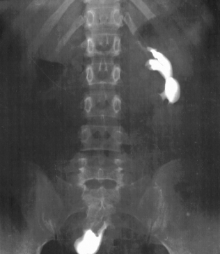
Physical examination revealed a palpable solid right adnexal mass with a smooth surface. Abnormal laboratory findings included only microscopic hematuria. Ultrasonography demonstrated an ectopic pelvic kidney with a small pelvic stone. This finding was confirmed by urography. The right kidney was associated with incomplete rotation (the renal pelvis was anterior instead of medial) and a short ureter. The left kidney showed excessive rotation (the renal pelvis lay laterally). The patient underwent successful extracorporeal shock wave lithotripsy.
When the mature kidney fails to reach its normal location, the condition is known as renal ectopia. An ectopic kidney can be found in one of the following positions: pelvic, iliac, abdominal, thoracic, or contralateral. Pelvic ectopia has been estimated to occur in 1 of 2100 to 3000 autopsies. The pelvic kidney is opposite the sacrum and below the aortic bifurcation. In some studies, ectopia and malrotation were inherited in screened families as an autosomal recessive trait, but this hypothesis needs further investigation.
Early in the embryonic life, during the end of the 5th week, the nephrogenic tissue is opposite the upper sacral somites. The process of kidney migration and rotation is completed by the end of the 8th week of gestation. Factors that may prevent the orderly movement of kidneys include ureteral bud maldevelopment, defective metanephric tissue that by itself fails to induce ascent, genetic abnormalities, maternal illnesses, or teratogenic causes.
The clinical significance of the ectopic kidney is that it is more prone to certain complications than are normally located kidneys. Ureteropelvic or ureterovesical junction obstruction and renal stones are more common. Pelvic ectopic kidneys are more liable to injury because they are unprotected by ribs and lie in a more anterior position. They may obstruct labor, although their surprising degree of mobility often allows them to be displaced into the lower portion of the abdomen and permit normal vaginal delivery.
The discrimination of a pathologic pelvic mass from an anatomic variation or unusual location of a normal organ can be a potential source of confusion and erroneous interpretation. Always consider a renal anomaly in the differential diagnosis of a “pelvic mass.”
FOR MORE INFORMATION:
Bauer SB. Anomalies of the upper urinary tract. In: Walsh PC, ed: Campbell’s Urology. Vol 3. Philadelphia: WB Saunders Company; 2002:1885-1924.