
Peer Reviewed
A Woman With Lobular Plaques on Her Shin
Authors:
Jason Le, DO
Naval Flight Surgeon for Command Training Air Wing 1 at Naval Air Station Meridian, Mississippi
Michael S. Dent, MD
Dermatology Clinic Department Head and Staff Dermatologist at Naval Hospital Pensacola, Florida
Disclosure:
The views expressed in this article are those of the authors and do not necessarily reflect the official policy or position of the Department of the Navy, the Department of Defense, or the US Government.
Citation:
Le J, Dent MS. A woman with lobular plaques on her shin. Consultant. 2019;59(2):53-54.
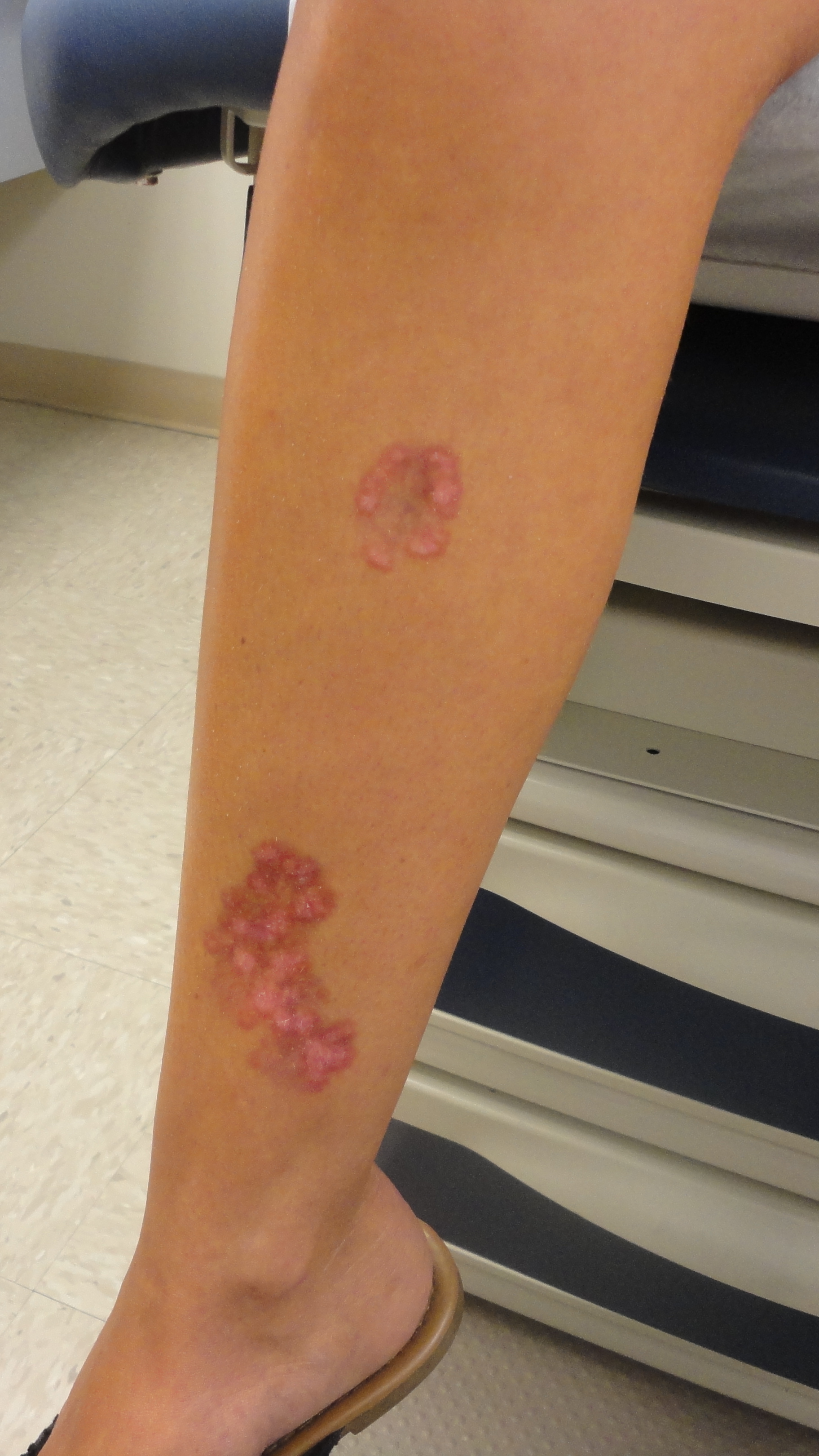
A 22-year-old woman with a history of type 1 diabetes mellitus presented with a several-year history of large asymptomatic plaques on her right shin that had been stable for the past 2 years. On physical examination, 2 well-demarcated, irregularly shaped, red, lobular plaques with atrophic, erythematous, and yellow-tinged portions were present on her right leg. The lesion on her upper right shin measured 3.5 × 3.0 cm, and the one on the lower right shin measured 7.0 × 4.0 cm (Figures).

Answer: Necrobiosis Lipoidica
A punch biopsy was done to confirm the diagnosis and to evaluate for other potential etiologies.
Necrobiosis lipoidica (NL), previously termed necrobiosis lipoidica diabeticorum due to its association with diabetes mellitus, usually type 1, is a rare chronic granulomatous skin disease.1-4 The condition presents predominantly in women and occurs at an average age of 25 years in persons with diabetes and 46 years in persons without diabetes.1,3,4 Associated conditions include sarcoidosis, inflammatory bowel disease, autoimmune thyroiditis, rheumatoid arthritis, and monoclonal gammopathy.3,4

Clinically, NL lesions are characterized by yellow-brown, atrophic, telangiectatic plaques with an elevated violaceous rim, located on the shins.1,3,4 The lesions are often bilateral and begin as small, firm, red-brown papules that gradually enlarge into plaques that atrophy and scar centrally.3,4 The lesions are often asymptomatic but may present with pruritus and/or pain, with ulcerations being a complication.1,4
On histopathology, the epidermis is often normal or atrophic with necrobiotic (connective tissue degeneration) collagen and palisading granulomas (histiocytes and lymphocytes surrounding a zone of altered collagen) in the dermis.1,2,4 The dermal interstitial infiltrate typically consists of histiocytes with many multinucleated giant cells, lymphocytes, and plasma cells.1,2,4
Differential Diagnosis
Granuloma annulare is a common, benign and self-limited skin disorder that more often occurs in women younger than 30 years. The condition often presents as asymptomatic, skin-colored to pink and pink-brown, arciform or annular plaques, with a predilection for the arms and hands. It can also involve the legs and feet and, less commonly, the trunk and face.1 The lesions are multivariate and include papular, subcutaneous, macular (or patch), and central perforating forms. On histopathology, the epidermis is normal; within the dermis (upper and middle), there are palisading granulomas surrounding a focus of mild necrobiosis and mucin accumulation, with perivascular lymphocytes often present (neutrophils or eosinophils are sometimes present).1,2
Sarcoidosis is a multisystem, granulomatous disease that affects individuals of all ages, sexes, and races, with a bimodal age distribution occurring between 25 to 35 years and 45 to 65 years.1 In the United States, the highest incidence is in African American women. Up to one-third of patients with systemic sarcoidosis present with cutaneous lesions as the first or only clinical manifestation.1 The cutaneous manifestations vary widely but commonly present as symmetrically distributed, red-brown to violaceous plaques and papules.1 Papules often occur in a perioral distribution, with plaques often developing on the trunk and extremities.1 Plaques can develop central, atrophic clearing and as such can present as annular lesions.1 On histopathology, the epidermis is usually normal with the presence of noncaseating (rarely caseating), well-demarcated, superficial and deep dermal epithelioid cell granulomas, often devoid of prominent infiltrates of lymphocytes or plasma cells (ie, a “naked granuloma”).1,2
Necrobiotic xanthogranuloma is a rare and progressive histiocytic cutaneous and subcutaneous disease, with multisystem involvement, affecting men and women equally and often occurring in persons approximately 60 years of age.1 The lesions present as asymptomatic indurated papules, nodules, or plaques with a yellow hue that may feature telangiectasias, atrophy, ulcerations, and scarring.1,2 Lesions often involve multiple sites on the face, trunk, and proximal extremities, with the periorbital regions most commonly affected and often with extracutaneous ocular manifestations.1 Most cases have an associated monoclonal gammopathy from plasma-cell dysplasia or lymphoproliferative disorders.1 Histopathology of nonulcerated lesions often demonstrates normal epidermis and superficial dermis with palisading xanthogranulomas found from the mid dermis extending into the subcutaneous tissues.1,2 The granulomas typically consist of histiocytes, foam cells, lymphoid follicles, plasma cells, and Touton giant cells with zones of necrobiosis containing cholesterol clefts.1,2
Diabetic dermopathy is an acquired hyperpigmentation disorder, usually in older persons with diabetes, occurring as pigmented pretibial patches.1,2 The lesions may begin as asymptomatic red to pink papules or plaques on the shins that evolve to brown atrophic macules and patches.1,2 Histopathology is nonspecific and may show epidermal edema, increased papillary dermal blood vessels, and/or perivascular lymphocytes.1,2
NL is associated with inconsistent treatment results and a lack of large randomized clinical trials.1,3,4 The serum glucose level does not have direct correlation with disease development or progression.1,3,4 Spontaneous remission has been reported in a low percentage of patients after 8 to 12 years of the condition.1,4
TREATMENT
Treatment considerations focus on complications arising from ulcerations, since the lesions are often difficult to heal and lead to bleeding and infection risks.1,3 Therefore, the most important treatment consideration is lifestyle modifications to minimize and avoid trauma to the affected area.1,3 Despite limited studies on efficacy, recommendations for first-line medical therapy are potent topical or intralesional corticosteroids in active borders of lesions, in order to halt disease progression by limiting inflammation and formation of granulation tissue.1,3 Topical and intralesional corticosteroids should be avoided in atrophic areas due to their potential for worsening of atrophy, which can lead to ulcerations.1,3,4
Other treatment modalities, such as phototherapy, biologics, and systemic immunomodulators, have been described mostly in case reports, prospective studies, and retrospective studies, with varying efficacies and remission rates. Phototherapy was the second-most cited therapy, with psoralen plus ultraviolet A (PUVA) therapy being the most widely used compared with photodynamic and ultraviolet A therapy.3,5 While phototherapy is generally safe with limited adverse effects, the largest disadvantage is the number of sessions required to achieve the desired effects, with 1 study showing an average of 47 PUVA sessions for remission.3 Biologics, such as infliximab, adalimumab, and etanercept, exert their effects as tumor necrosis factor α inhibitors, which play a key role in formation of granulomas, but recurrences were common after discontinuation of therapy.3,4,5 Systemic immunotherapy such as cyclosporine, fumaric acid, and mycophenolate mofetil were commonly cited, and similar to biologics, NL tends to recur after discontinuation of therapy.3,5,6
Surgical therapy is reserved for severe and refractory ulcerations but may require excision deep into the fascia to prevent recurrence and split-thickness graft for wound healing.1,3 NL has a tendency to exhibit koebnerization, which presents challenges to surgical treatments.3,4
Our patient’s lesions have been stable and asymptomatic, have not developed ulcerations or secondary infections, and present minimal bleeding risk. We are monitoring for now and may consider treatment as needed for any progression of disease.
References:
- Rosenbach MA, Wanat KA, Reisenauer A, White KP, Korcheva V, White CR Jr. Non-infectious granulomas. In: Bolognia JL, Schaffer JB, Cerroni L, eds. Dermatology. Vol 2. 4th ed. Philadelphia, PA: Elsevier; 2017:1644-1663.
- Rapini RP. Non-infectious granulomas. In: Rapini R. Practical Dermatopathology. 2nd ed. Philadelphia, PA: Elsevier Saunders; 2012:101-118.
- Reid SD, Ladizinski B, Lee K, Baibergenova A, Alavi A. Update on necrobiosis lipoidica: a review of etiology, diagnosis, and treatment options. J Am Acad Dermatol. 2013;69(5):783-791.
- Lepe K, Salazar FJ. Necrobiosis lipoidica. StatPearls. https://www.ncbi.nlm.nih.gov/books/NBK459318/. Updated October 27, 2018. Accessed January 17, 2019.
- Peckruhn M, Tittelbach J, Elsner P. Updated: treatment of necrobiosis lipoidica. J Dtsch Dermatol Ges. 2017;15(2):151-157.
- Feily A, Mehraban S. Treatment modalities of necrobiosis lipoidica: a concise systematic review. Dermatol Reports. 2015;7(2):5749.