Peer Reviewed
A Recurring Rash on an Older Man’s Thigh
Correct answer: C. Fixed drug eruption
A fixed drug eruption was diagnosed. The patient was subsequently told to stop taking doxycycline and was initiated on topical clindamycin solution 1% applied to the intertriginous areas once a day for 2 weeks to treat the hidradenitis suppurativa.
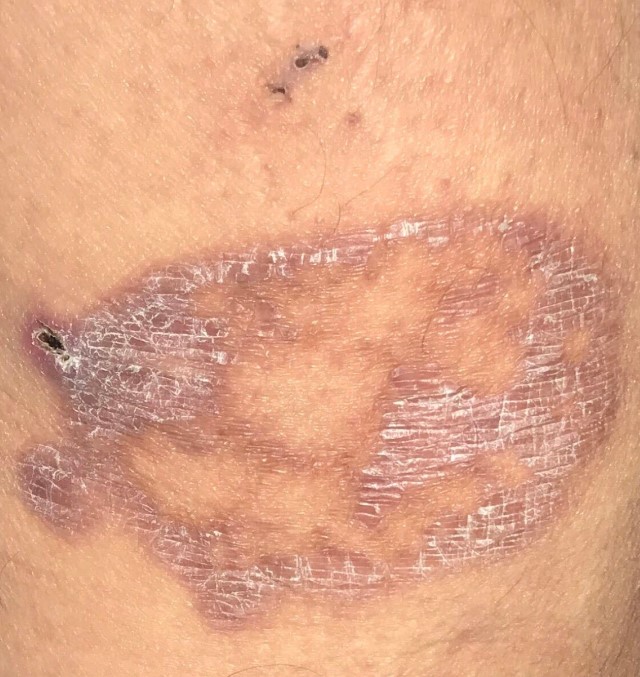
Discussion. Fixed drug eruptions are cutaneous drug reactions characterized by recurrent skin lesions that occur in the same location upon exposure to the offending drug. While many drugs have been reported to cause fixed drug eruptions, the most common culprits are nonsteroidal anti-inflammatory drugs and a variety of antibiotics, such as amoxicillin, fluoroquinolones, and doxycycline.1 Lesions are usually solitary and typically present as well-demarcated, oval to round, dusky erythematous plaques that may vesiculate or blister and often heal with residual hyperpigmentation.2
Symptoms such as pruritus or pain are occasionally reported, but systemic symptoms are usually absent. Sites of predilection for fixed drug eruptions include the genitalia, lips, perianal area, and distal extremities.2 Histologically, fixed drug eruptions typically show interface dermatitis, pigmentary incontinence with civatte bodies, and lymphocytic infiltration of the dermis with scattered eosinophils (Figure 2).3 Eruptions usually occur within minutes to several hours after exposure to the offending drug and resolve after 1 to 2 weeks of discontinuation, with ensuing post-inflammatory hyperpigmentation. Discontinuation of the offending drug is the most important aspect of treatment. Symptomatic therapy includes oral H1 antihistamine and a 7- to 10-day course of topical corticosteroids.1,2

Figure 2. A hematoxylin-eosin stain was conducted for our patient. At 20× magnification, results showed basket-weave stratum corneum and a vacuolar interface obscuring the dermoepidermal junction. The arrow indicates a civatte body.The deep variant of erythema annulare centrifugum, which is also associated with medication exposure, can be considered in the differential diagnosis for this patient.3 However, erythema annulare centrifugum typically presents as a solitary, annular, erythematous patch with an edematous leading edge but minimal scale. Erythema annulare centrifugum usually has a waxing and waning clinical course over months prior to spontaneous resolution.3,4
Granuloma annulare is another differential diagnosis to consider, because its lesions also appear smooth and annular. However, granuloma annulare typically lacks the waxing and waning pattern observed with our patient’s lesion. In addition, granuloma annulare can be differentiated histologically by its characteristic lymphohistocytic infiltrate, collagen degeneration, and mucin deposition, which are absent in fixed drug eruptions.5
Necrobiosis lipoidica is an uncommon, granulomatous disease that typically presents as violaceous papules or nodules that progress to dark yellow plaques that flatten and become atrophic with time. Erosion and ulceration are common complications. While there is a strong association with type 2 diabetes, this association is not ubiquitous.6 Unlike fixed drug eruptions, necrobiosis lipoidica most commonly presents on the lower extremities and typically has a chronic and progressive course.7 Histologically, necrobiosis lipoidica shows palisaded arrangement and granulomatous dermatitis with an abundance of histiocytes and multinucleated giant cells.8
Majocchi granuloma is a dermatophyte infection that is most commonly caused by Trichophyton rubrum.9 Unlike most other dermatophyte infections, Majocchi granuloma involves the dermis and subcutaneous tissue. Clinically, Majocchi granuloma presents with an irregular scaly plaque with an erythematous base. Scattered suppurative follicular papules and pustules on the plaques are common findings. Topical antifungal treatment is insufficient; treatment of Majocchi granuloma requires oral antifungal therapy such as terbinafine, griseofulvin, or itraconazole.9
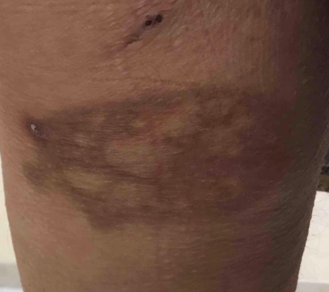
Patient outcome. Fixed drug eruptions are usually benign and self-resolving. Our patient's rash on the left posterior thigh began resolving on day 5 after discontinuation of oral doxycycline with marked reduction in pain, pruritis, and erythema (Figure 3). By 2 months after discontinuation of oral doxycycline, all symptoms completely resolved with post-Inflammatory hyperpigmentation present at the site of the rash (Figure 4).
Figure 3. After 5 days of discontinuing doxycycline, the lesion started to heal. Note the double open comedones, which are a classic sign of hidradenitis suppurativa.
Figure 4. After 2 months of discontinuing doxycycline, the lesion healed with residual post-inflammatory hyperpigmentation.References
1. Ben Fadhel N, Chaabane A, Ammar H, et al. Clinical features, culprit drugs, and allergology workup in 41 cases of fixed drug eruption. Contact Dermatitis. 2019;81(5):336-340. https://doi.org/10.1111/cod.13351
2. Flowers H, Brodell R, Brents M, Wyatt JP. Fixed drug eruptions: presentation, diagnosis, and management. South Med J. 2014;107(11):724-727. https://doi.org/10.14423/smj.0000000000000195
3. Kim DH, Lee JH, Lee JY, Park YM. Erythema annulare centrifugum: analysis of associated diseases and clinical outcomes according to histopathologic classification. Ann Dermatol. 2016;28(2):257-259. https://doi.org/10.5021/ad.2016.28.2.257
4. Kim KJ, Chang SE, Choi JH, Sung KJ, Moon KC, Koh JK. Clinicopathologic analysis of 66 cases of erythema annulare centrifugum. J Dermatol. 2002;29(2):61-67. https://doi.org/10.1111/j.1346-8138.2002.tb00167.x
5. Dabski K, Winkelmann RK. Generalized granuloma annulare: histopathology and immunopathology. Systematic review of 100 cases and comparison with localized granuloma annulare. J Am Acad Dermatol. 1989;20(1):28-39. https://doi.org/10.1016/s0190-9622(89)70004-9
6. Erfurt-Berge C, Dissemond J, Schwede K, et al. Updated results of 100 patients on clinical features and therapeutic options in necrobiosis lipoidica in a retrospective multicentre study. Eur J Dermatol. 2015;25(6):595-601. https://doi.org/10.1684/ejd.2015.2636
7. Jockenhöfer F, Kröger K, Klode J, Renner R, Erfurt-Berge C, Dissemond J. Cofactors and comorbidities of necrobiosis lipoidica: analysis of the German DRG data from 2012. J Dtsch Dermatol Ges. 2016;14(3):277-284. https://doi.org/10.1111/ddg.12749
8. Gibson LE, Reizner GT, Winkelmann RK. Necrobiosis lipoidica diabeticorum with cholesterol clefts in the differential diagnosis of necrobiotic xanthogranuloma. J Cutan Pathol. 1988;15(1):18-21. https://doi.org/10.1111/j.1600-0560.1988.tb00509.x
9. Ilkit M, Durdu M, Karakaş M. Majocchi's granuloma: a symptom complex caused by fungal pathogens. Med Mycol. 2012;50(5):449-457. https://doi.org/10.3109/13693786.2012.669503