
Peer Reviewed
What Is Causing the Twinkle in a Patient’s Eye?
AUTHORS:
Joseph T. Garand, MD1 • Adam Gordon, OD, MPH2 • Seth F. Patterson, MD1
AFFILIATIONS:
1Prisma Health Greer Family Medicine Residency Program, Department of Family Medicine, University of South Carolina School of Medicine Greenville, South Carolina
2University of Alabama at Birmingham School of Optometry, Birmingham, Alabama
CITATION:
Garand JT, Gordon A, Patterson SF. What is causing the twinkle in a patient’s eye? Consultant. 2020;50(5):e10. doi:10.25270/con.2020.03.00019
Received January 25, 2020. Accepted February 21, 2020.
CORRESPONDENCE:
Joseph T. Garand, MD, Medical Director, Prisma Health System Greer Family Medicine Residency Program, 109 Physicians Dr Ste A, Greer, SC 29650 (joseph.garand@prismahealth.org)
DISCLOSURES:
The authors report no relevant financial relationships.
A 68-year-old new patient presented to a primary care provider for a routine checkup. Examination of the left eye with an external light source (otoscope) revealed the findings depicted in Figure 1.

Figure 1. The patient’s left eye during examination with an otoscope light source.
ANSWER: PSEUDOPHAKIA
The twinkle in this patient’s eye is a result of pseudophakia, with a normal Purkinje 1 (P1) reflex and prominent Purkinje 3 and 4 (P3 and P4) images (reflexes) (Figure 2). Pseudophakia refers to an eye in which the natural lens has been replaced with an implanted artificial intraocular lens (IOL).

Figure 2. The patient’s left eye during examination with an otoscope light source showing a normal P1 image and prominent P3 and P4 images.
Very little has been published in the primary care literature on the Purkinje reflexes, which are most commonly of interest to ophthalmologists in patients who have had their natural lens replaced with an IOL. Because ophthalmologists can directly visualize the Purkinje reflexes in the operating room using a surgical microscope, the now readily visualized Purkinje reflexes are sidelined as findings of little importance.1
Purkinje reflexes are reflections from the cornea and lens of the eye.2 They consist of the corneal reflex (P1) and other Purkinje images (P2, P3, and P4). At least 3 of the 4 possible Purkinje images are usually visible with the use of a slit lamp. P1 is the reflection from the anterior surface of the cornea and can be seen in all eyes as long as the cornea is clear. It is the brightest, moves the slowest, parallels the movement of light source, and is on the same side of the cornea as the light source. P2 is the reflection from the posterior surface of the cornea and can only be seen with a slit lamp. P3 is the reflection from the anterior surface of the lens; it is usually the largest, moves the fastest, and parallels movement of the light source. P4 is the reflection from the posterior surface of the lens. It is mid-sized and moves at moderate speed, but it moves in a direction opposite of the light source and thus opposite in direction to P1. P4 is a reflection from the concave posterior lens; thus it will be an inverted image of the light source (Figure 3).3

Figure 3. Purkinje images. (Wikimedia Commons, https://commons.wikimedia.org/wiki/File:Diagram_of_four_Purkinje_images.svg)
P1 is the only light reflex that the primary care provider will typically observe with a penlight or other external light source in a phakic (containing a natural lens) eye. The other Purkinje images are not typically seen in a phakic eye unless a slit lamp is utilized. In the pseudophakic eye, however, the refracting surfaces of the intraocular lens (IOL) have excellent optical properties. IOLs made of high-index acrylic can have a relative glare intensity of reflections in the range of several hundred to several thousand times greater than the natural lens.4 Because of these properties, P3 and P4 are now readily observed as rounded shining reflections. The intensity of the P3 with IOLs is why some patients report having a “glimmer” in their eyes after IOL implantation.3
HOW TO MAKE THE DIAGNOSIS
The technique for observing Purkinje reflexes in a pseudophakic eye is as follows: The examiner, facing the patient, shines the light source on the lateral aspect of the patient’s eye. The corneal reflex (P1) is visualized on the lateral aspect of the pupil. The light source is then moved medially as the examiner looks for additional Purkinje light reflexes approaching from the medial aspect of the pupil. P3 and P4 in this case look like small shining bubbles (Figure 1) as they take on the characteristic of the otoscope light source (Figure 4). The accompanying Video demonstrates visualization of the Purkinje reflexes in a patient with an IOL implant.
Video. Visualization of the Purkinje reflexes in a patient with an IOL implant.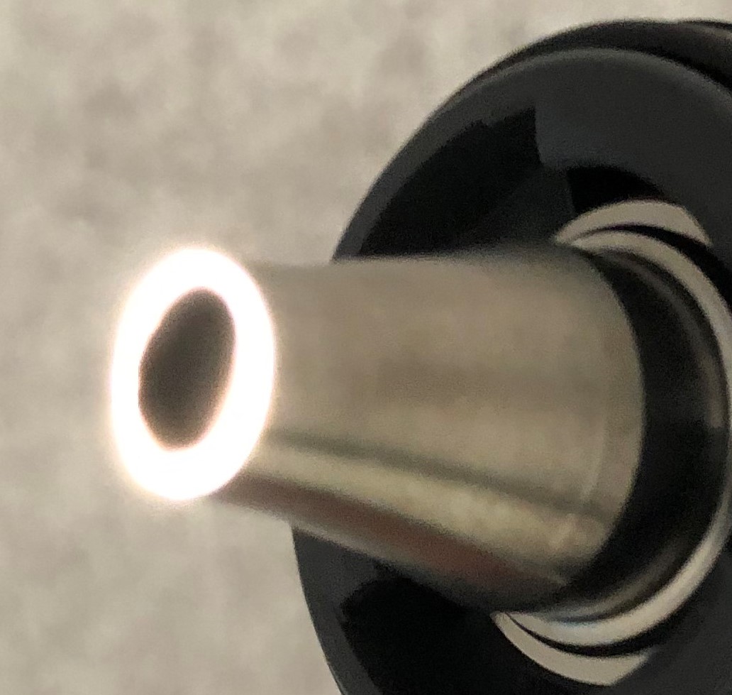
Figure 4. An illuminated otoscope. Note in Figure 1 and in the accompanying Video, the halo appearances of P3 and P4 are due to the halo of light projecting from the otoscope.
Even experienced primary care providers often do not recognize or understand this rapid and simple way to detect pseudophakia. Given that many patients do not recall the specifics of their histories, this examination finding can help clinicians better document the patient’s ophthalmologic history.
REFERENCES:
- Gellrich M-M, Kandzia C. Purkinje images in slit lamp videography: video article [in German]. Ophthalmologe. 2016;113(9):789-793. doi:10.1007/s00347-016-0343-4
- Chang DH, Waring GO IV. The subject-fixated coaxially sighted corneal light reflex: a clinical marker for centration of refractive treatments and devices. Am J Ophthalmol. 2014;158(5):863-874.e2. doi:10.1016/j.ajo.2014.06.028
- Cavero I, Guillon J-M, Holzgrefe HH. Reminiscing about Jan Evangelista Purkinje: a pioneer of modern experimental physiology. Adv Physiol Educ. 2017;41:528-538. doi:10.1152/advan.00068.2017.5281043
- Chang DH. Characterizing IOLs with Purkinje images. Cataract & Refractive Surgery Today. https://crstoday.com/articles/2011-oct/characterizing-iols-with-purkinje-images/. Published October 2011. Accessed February 24, 2020.