
Authors:
Gina Duong, BA
Medical Student, McGovern Medical School at the University of Texas Health Science Center at Houston
LaTanya Love, MD
Associate Professor of Pediatrics, McGovern Medical School at the University of Texas Health Science Center at Houston
Citation:
Duong G, Love L. Bilateral dental lamina cysts. Consultant. 2019;59(2):58-59.
An 8-day-old boy presented to our clinic for newborn hospital follow-up. The patient had been born at 37 weeks and 3 days via spontaneous vaginal delivery to a gravida 1, para 0 mother who had had good prenatal care and who had experienced no complications of pregnancy or labor. The patient was feeding, voiding, and stooling without problems.
On physical examination, the neonate was found to have small, smooth cystic masses of approximately 1 cm in diameter located on the crest of the alveolus, where the canine adult tooth eventually erupts (Figure 1).

Figure 1.
These masses were noted on the bilateral mandibular alveolar ridges symmetrically, as well as on the bilateral maxillary alveolar ridges, also symmetrically (Figure 2).
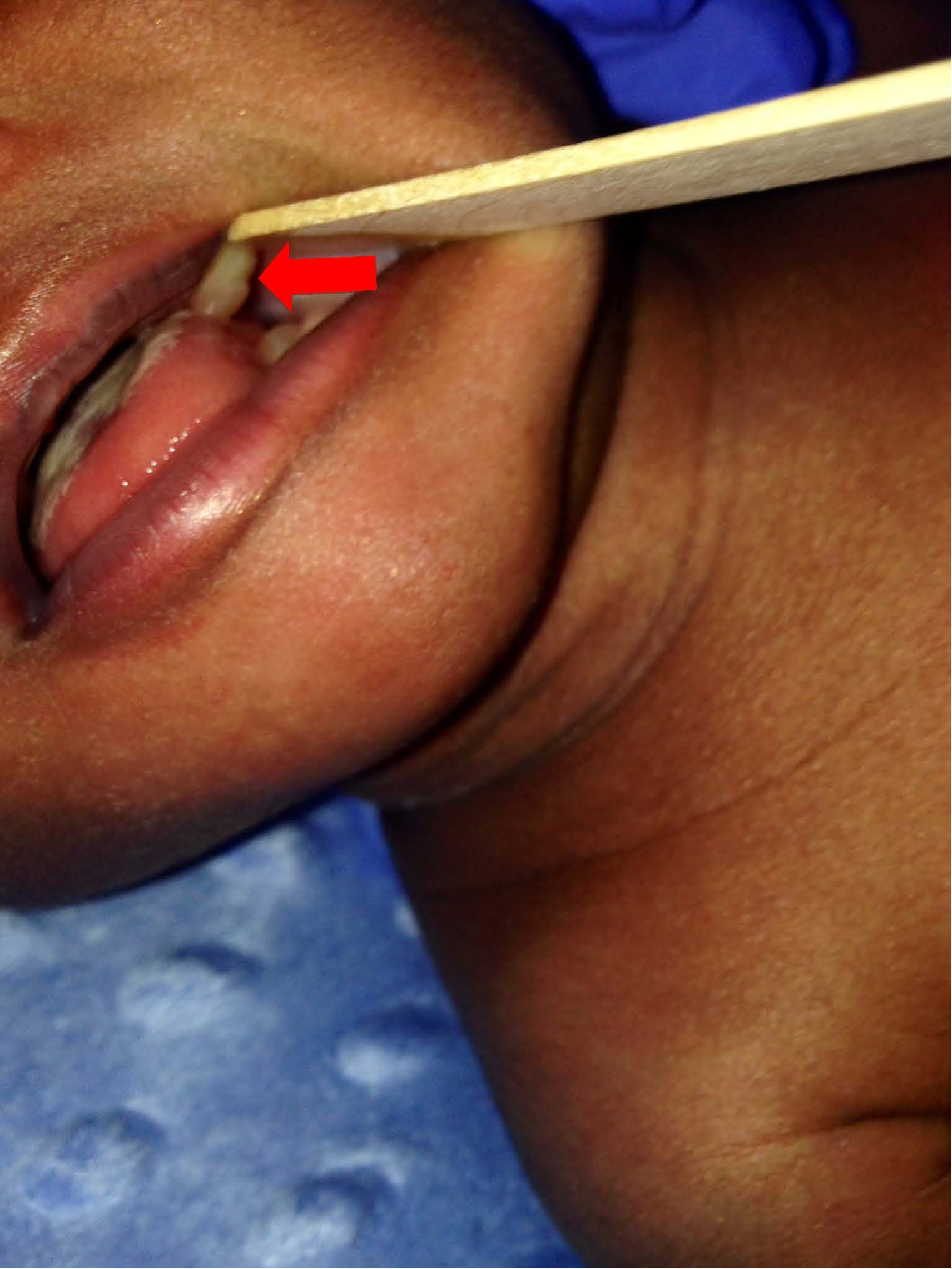
Figure 2.
These lesions did not appear to be tender to palpation, and they did not interfere with feeding. Also of note, the neonate had a mild case of oral thrush, which was most apparent on the tongue (Figure 3).
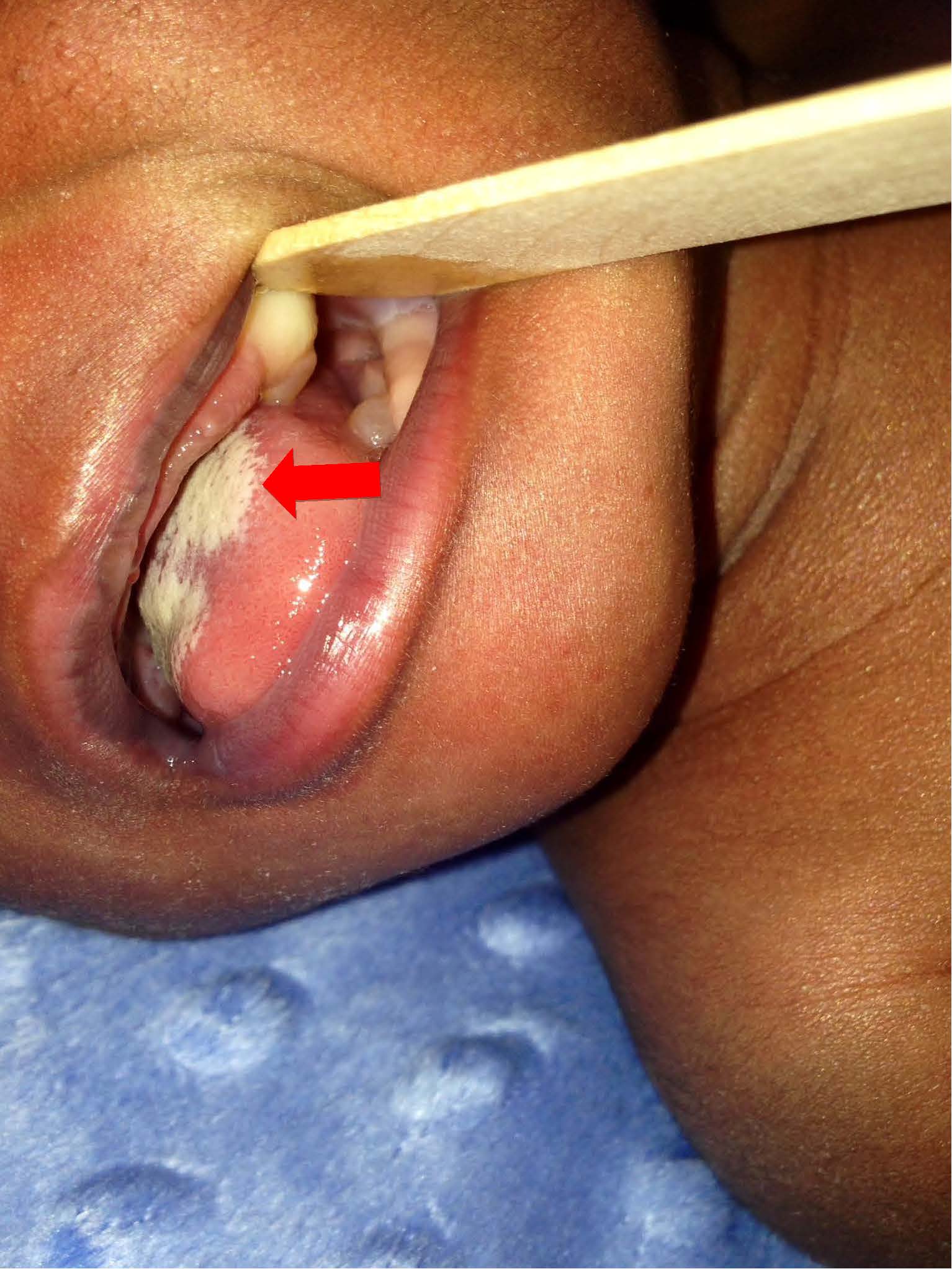
Figure 3.
Discussion
Oral soft tissue lesions in newborns can be a concern for parents. Most oral soft tissue lesions found before 6 months of age are benign and self-limiting. Differential diagnosis includes dental laminal cysts, Bohn nodules, Epstein pearls, natal teeth, neonatal teeth, and congenital epulis. Patients of all ages are susceptible to oral infections, which also can present as mass lesions in the oral cavity. Therefore, infectious causes such as herpes simplex virus and human papillomavirus should be considered in the differential diagnosis for any patient with an oral lesion.
George and colleagues in 2008 conducted a study of 1038 newborns in India, reporting the prevalence of dental lamina cysts at birth as 13.8%, Epstein pearls as 35.2%, and Bohn nodules as 47.4%.1 The classic categorization of the cystic oral lesions dates to 1967, when Fromm categorized them based on histology, tissue of origin, and location.2 These 3 cystic oral lesions are keratin-filled nodules, typically millimeters in size, but they have different proposed tissues of origin.2
Dental lamina cysts arise from the remnants of the dental lamina, which is the ectodermal band giving rise to dentition. They most often present as multiple white, yellow, or pink nodules on the alveolar ridge in newborns, frequently causing them to be mistaken for neonatal or natal teeth.3 Histologically, they are true cysts with an epithelial lining and are filled with keratin and occasionally inflammatory cells.4 They are asymptomatic and rarely cause discomfort to the patient.5 After birth, dental lamina cysts often degenerate, starting with atrophy and resorption of the keratin and epithelial cells inside the cyst. Then, some cysts degenerate and get digested by giant cells, whereas some open onto the surface leaving clefts or become involved in developing teeth.6
Bohn nodules are thought to arise from the dental lamina or from minor salivary glands. They present as white, yellow, or pink nodules at the buccal or lingual aspects of the alveolar ridges. It has been noted that dental lamina cysts and Bohn nodules are histologically the same and have the same in origin; therefore, they should be categorized together and called gingival cysts.7
Epstein pearls, also called palatine raphe cysts of the newborn, arise from the epithelial remnants along the fusion line of the palatal folds and the nasal septum. They present as white or yellow nodules at the median palatine raphe, most commonly at the junction of the soft and hard palate.7
The diagnosis of cystic oral lesions is made clinically. Because of their benign nature, biopsies are not routinely performed. They are self-limiting, commonly spontaneously breaking, degenerating, or reducing in size within 2 weeks to 5 months of postnatal life. They do not warrant any treatment.4
Congenital epulides, or congenital granular cell lesions, are rare granular cell hamartomas in newborns that most commonly present as smooth masses on the maxillary alveolar process.8 They often regress spontaneously in 3 to 6 months. However, unlike cystic oral lesions, they can measure up to a few centimeters in diameter, causing dyspnea, coughing, difficulty suckling, and difficulty swallowing. Therefore, surgical removal may be necessary. Perinatal magnetic resonance imaging can help distinguish this mass from the cystic oral lesions and help in surgical planning.9
The patient in this case had multiple 1-cm lesions that were smooth and cystic on his mandibular and maxillary alveolar processes. Due to the age of the patient, the location of the lesions, and the relative incidence of dental lamina cysts compared with other lesions, dental lamina cysts were highest on the differential diagnosis list. Although dental lamina cysts can present as solitary lesions, they presented as clusters of lesions in this patient.
The family of the patient in this case was reassured of the self-limiting and benign nature of the patient’s dental lamina cysts. The cysts were no longer detected at the 6-month well child visit.
References:
- George D, Bhat SS, Hegde SK. Oral findings in newborn children in and around Mangalore, Karnataka State, India. Med Princ Pract. 2008;17(5):385-389.
- Fromm A. Epstein pearls, Bohn’s nodules and inclusion-cysts of the oral cavity. J Dent Child. 1967;34(4):275-287.
- Bodenhoff J, Gorlin R. Natal and neonatal teeth: folklore and fact. Pediatrics. 1963;32(6):1087-109
- Marini R, Chipaila N, Monaco A, Vitolo D, Sfasciotti GL. Unusual symptomatic inclusion cysts in a newborn: a case report. J Med Case Rep. 2014;8:31
- Benni DB, Sirur D. Gingival cyst of the newborn: a case report. Int Dent. 2013;3(2):32-34.
- Singh RK, Kumar R, Pandey RK, Singh K. Dental lamina cysts in a newborn infant. BMJ Case Rep. 2012;2012:bcr2012007061.
- Van Heerden WFP, Van Zyl AW. Diagnosis and management of oral lesions and conditions in the newborn. S Afr Fam Pract. 2010;52(6):489-491.
- Mabongo M, Wood NH, Lemmer J, Feller, L. Congenital epulis: a case report. SADJ. 2008;63(6):350-351.
- Kumar RM, Bavle RM, Umashankar DN, Sharma R. Congenital epulis of the newborn. J Oral Maxillofac Pathol. 2015;19(3):407.