
Peer Reviewed
Neonatal Hemophilia A
AUTHORS:
Surasak Puvabanditsin, MD1 • Rannan Kased, DO1 • Akreeti Maskey, MD1 • Rajeev Mehta, MD1 • Susan Murphy, MD2,3
AFFILIATIONS:
1Department of Pediatrics, Rutgers Robert Wood Johnson Medical School, New Brunswick, New Jersey
2Director of Pediatric Thrombosis and Hemostasis, Rutgers Robert Wood Johnson Medical School, New Brunswick, New Jersey
3Pediatric Hematologist/Oncologist, Rutgers Robert Wood Johnson Medical School, New Brunswick, New Jersey
CITATION:
Puvabanditsin S, Kased R, Maskey A, Mehta R, Murphy S. Neonatal hemophilia A. Consultant. 2022;62(2):e28-e30. doi:10.25270/con.2021.05.00006
Received October 10, 2020. Accepted January 19, 2021. Published online May 21, 2021.
DISCLOSURES:
The authors report no relevant financial relationships.
CORRESPONDENCE:
Surasak Puvabanditsin, MD, Rutgers Robert Wood Johnson Medical School, 1 Robert Wood Johnson Place, New Brunswick, NJ 08903 (surasak1@aol.com)
A 2-day-old boy was brought to the well-baby nursery with edema of the right thigh. There was no abnormality noted at birth.
History. The patient was born at 38 weeks of gestation without complications after an uncomplicated pregnancy to a primigravida who was 31 years of age at the time of birth. The patient’s birth weight was 2.87 kg. His mother was a hepatitis B virus carrier, therefore the patient had received hepatitis B immunoglobulin and vaccine, as well as vitamin K1 intramuscularly on his thighs, soon after birth. There was no family history of genetic diseases, and his prenatal history was unremarkable.
Physical examination. On physical examination, the infant appeared well and interactive. He was normocephalic, with normally set ears and without dysmorphic facial features. His lungs were clear to auscultation, with symmetric chest wall expansion. A cardiovascular examination revealed a normal heart rate, with regular rhythm and without murmurs. His abdomen was soft, nontender, and nondistended. There was no organomegaly. He had normal genitalia for his age and sex. He had normal range of motion, with normal strength and no head lag. He responded to stimulation, had a symmetric Moro reflex, positive suck reflex, and positive palmar grasp reflex.
The skin over the swollen right thigh had ecchymosis and discoloration (Figures 1 and 2). The infant was transferred to the neonatal intensive care unit (NICU) nursery. A blood culture was obtained, and antibiotics were initiated. Peripheral intravenous (IV) fluid was infused on the left forearm and hand. On the day after NICU admission (day 3 of the infant’s life), ecchymosis of his left hand and forearm were noted after removal of the IV angiocatheter (Figure 3). The bleeding at the IV site was noted to prolong. Coagulation (ie, activated partial thromboplastintime [aPTT] and prothrombin time) and platelet count studies were obtained. The aPTT was prolonged. The diagnosis of hemophilia was made after the factor VIII level confirmed the diagnosis.
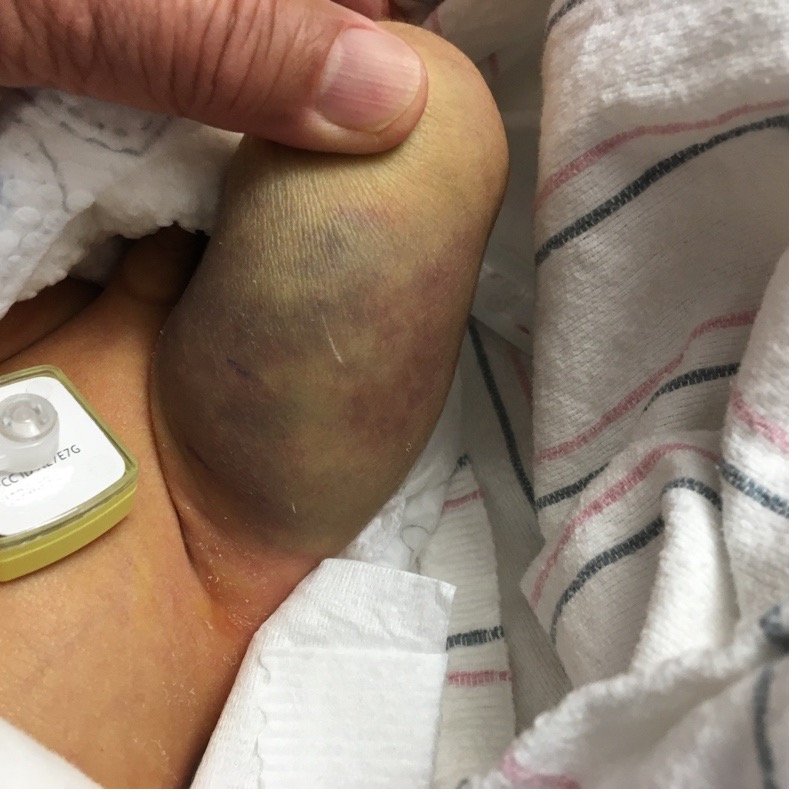
Figure 1. Swelling and ecchymosis of the neonate’s left thigh.
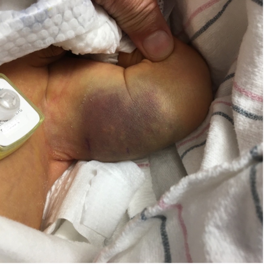
Figure 2. Swelling and ecchymosis of the neonate’s left thigh.
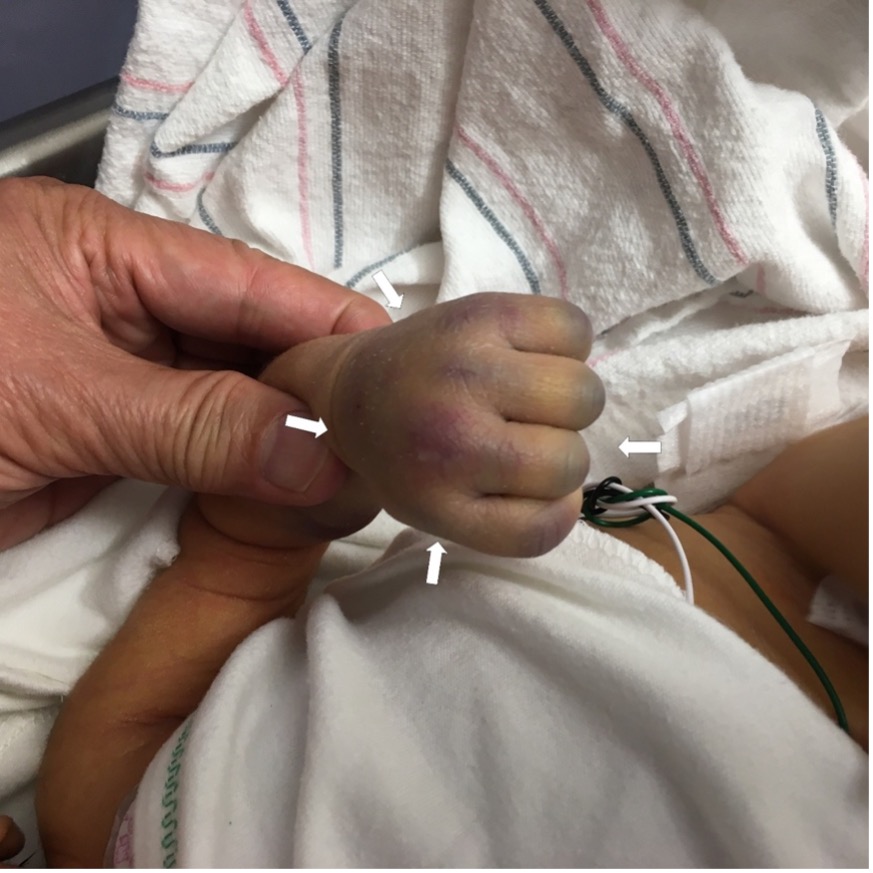
Figure 3. Ecchymosis of the neonate’s left hand and fingers where the angiocatheter was indwelled.
Treatment. The patient received factor VIII concentrates. The ecchymosis and swelling gradually resolved without any complications. He was discharged home on day 10 of life. Within a few days of discharge, he was seen by our pediatric hematologist at the clinic where his parents were counseled about potential problems (eg, heel sticks, vaccinations, intramuscular injection), including those that may relate to major bleeding, such as intracranial hemorrhage (ICH).
Discussion. Hemophilia A is an inherited bleeding disorder caused by deficiency of coagulation factor VIII. Hemophilia A affects more than 1.2 million individuals (mostly boys and men) worldwide. Hemophilia occurs in all ethnic groups and throughout the world. Hemophilia A occurs in approximately 1 in every 4000 to 1 in every 5000 live male births.1 It is transmitted in an X-linked recessive pattern. Transmission is from female carriers to male children. Approximately half of male children of a female carrier will be affected. Female children of affected males are obligate carriers. Father-to-son transmission does not occur. Male children within a family who inherit the familial mutation will all have approximately the same degree of factor deficiency and similar severity of disease because they share the same genetic defect.
A large proportion of affected individuals have a negative family history, and a negative family history cannot be used to exclude the possibility of hemophilia. A negative family history is typically explained by a de novo hemophilia mutation in the mother.2-4 Studies have demonstrated that sporadic causes account for as much as 45% of cases of severe hemophilia A.5
The range of ages for first bleed is wide, with some children having severe bleeding at birth and others who do not experience a bleeding episode until age 4 years.6,7 Common sites of bleeding in neonates include the central nervous system, extracranial sites such as cephalohematoma, and sites of medical interventions including circumcision, heel sticks, and venipunctures.8 The overall incidence of intracranial ICH in neonates is approximately 3.5% to 5.5% at birth.9 Routine cranial ultrasonography scans in all neonates before discharge have been recommended in some studies.10,11 Cranial magnetic resonance imaging and computed tomography (CT) scanning should be performed in symptomatic neonates with ICH. Our patient’s neurosonogram and cranial CT scans were normal.
At birth, a diagnosis of hemophilia can be made using cord blood or a venous blood sample.12 Cord blood testing is preferred, if possible, to avoid the risk of bleeding with venipuncture.13 Cord blood is tested for factor activity level of the appropriate factor. The diagnosis of hemophilia A (inherited factor VIII deficiency) requires confirmation of a factor VIII activity level lower than 40% of normal (< 0.40 IU/mL).14,15
Our patient received 0.5 mL of the hepatitis B virus vaccine and 0.1 mL of vitamin K1 intramuscularly on the right thigh, and 0.5 mL of hepatitis B immunoglobulin was administered on the left thigh. The swelling of the right thigh was apparent at about 36 hours after the intramuscular injections.
The differential diagnosis of hemophilia includes von Willebrand disease, inherited platelet disorders, factor XI deficiency and factor XIII deficiency. Nicolau syndrome, or embolia cutis medicamentosa, is another condition that would be considered in the differential diagnosis as a complication of intramuscular injection in neonates. Nicolau syndrome is a rare cutaneous drug reaction that occurs at the site of an intramuscular drug injection. It was first described in 1924 by Freudenthal in patients treated with bismuth salts for syphilis.16 Nicolau syndrome has been reported with the administration of various other drugs and vaccines, such as vitamin K, penicillins, local anesthetics, corticosteroids, nonsteroidal anti-inflammatory drugs, and the diphtheria-tetanus-pertussis vaccine.17,18,19 The proposed pathogenesis is direct trauma or irritation of the vascular structures with a compression by the arterial embolism of the drug itself and crystallization of aqueous drugs in the vessels.
References
1. Iorio A, Stonebraker JS, Chambost H, et al; Data and Demographics Committee of the World Federation of Hemophilia. Establishing the prevalence and prevalence at birth of hemophilia in males: a meta-analytic approach using national registries. Ann Intern Med. 2019;171(8):540-546. https://doi.org/10.7326/M19-1208
2. Thompson AR, Bajaj SP, Chen SH, MacGillivray RT. "Founder" effect in different families with haemophilia B mutation. Lancet. 1990;335(8686):418. https://doi.org/10.1016/0140-6736(90)90259-8
3. Lawn RM. The molecular genetics of hemophilia: blood clotting factors VIII and IX. Cell. 1985;42(2):405-406. https://doi.org/10.1016/0092-8674(85)90094-7
4. Carcao MD. The diagnosis and management of congenital hemophilia. Semin Thromb Hemost. 2012;38(7):727-747. https://doi.org/10.1055/s-0032-1326786
5. Kasper CK, Lin JC. Prevalence of sporadic and familial haemophilia. Haemophilia. 2007;13(1):90-92. https://doi.org/10.1111/j.1365-2516.2006.01397.x
6. Escuriola Ettingshausen C, Halimeh S, Kurnik K, et al. Symptomatic onset of severe hemophilia A in childhood is dependent on the presence of prothrombotic risk factors. Thromb Haemost. 2001;85(2):218. http://dx.doi.org/10.1007/978-3-642-59383-3_9
7. Pollmann H, Richter H, Ringkamp H, Jürgens H. When are children diagnosed as having severe haemophilia and when do they start to bleed? A 10-year single-centre PUP study. Eur J Pediatr. 1999;158(suppl 3):S166-S170. https://doi.org/10.1007/pl00014347
8. Kulkarni R, Presley RJ, Lusher JM, et al. Complications of haemophilia in babies (first two years of life): a report from the Centers for Disease Control and Prevention Universal Data Collection System. Haemophilia. 2017;23(2):207-214. https://doi.org/10.1111/hae.13081
9. Richards M, Lavigne Lissalde G, Combescure C, et al. Neonatal bleeding in haemophilia: a European cohort study. Br J Haematol. 2012;156(3):374-382. https://doi.org/10.1111/j.1365-2141.2011.08967.x
10. Chalmers EA, Williams MD, Richards M, et al. Management of neonates with inherited bleeding disorders--a survey of current UK practice. Haemophilia. 2005;11(2):186-187. https://doi.org/10.1111/j.1365-2516.2005.01072.x
11. Williams MD, Chalmers EA, Gibson BE; Haemostasis and Thrombosis Task Force, British Committee for Standards in Haematology. The investigation and management of neonatal haemostasis and thrombosis. Br J Haematol. 2002;119(2):295-309. https://doi.org/10.1046/j.1365-2141.2002.03674.x
12. Price VE, Hawes SA, Chan AK. A practical approach to hemophilia care in children. Paediatr Child Health. 2007;12(5):381-383. https://doi.org/10.1093/pch/12.5.381
13. National Hemophilia Foundation. MASAC Document 265 - MASAC Guidelines for Pregnancy and Perinatal Management of Women with Inherited Bleeding Disorders and Carriers of Hemophilia A or B. Published March 4, 2021. Accessed May 5, 2021. https://www.hemophilia.org/healthcare-professionals/guidelines-on-care/masac-documents/masac-document-265-masac-guidelines-for-pregnancy-and-perinatal-management-of-women-with-inherited-bleeding-disorders-and-carriers-of-hemophilia-a-or-b
14. Makris M, Oldenburg J, Mauser-Bunschoten EP, et al. The definition, diagnosis and management of mild hemophilia A: communication from the SSC of the ISTH. J Thromb Haemost. 2018;16(12):2530-2533. https://doi.org/10.1111/jth.14315
15. White GC 2nd, Rosendaal F, Aledort LM, et al. Definitions in hemophilia: Recommendation of the scientific subcommittee on factor VIII and factor IX of the scientific and standardization committee of the International Society on Thrombosis and Haemostasis. Thromb Haemost. 2001;85(3):560. http://dx.doi.org/10.1055/s-0037-1615621
16. Freudenthal W. Lokales embolisches Bismogenol-Exanthem. Arch f Dermat. 1924;147:155-160. https://doi.org/10.1007/BF01828197
17. Puvabanditsin S, Garrow E, Weerasethsiri R, Joshi M, Brandsma E. Nicolau's syndrome induced by intramuscular vitamin K injection in two extremely low birth weight infants. Int J Dermatol. 2010;49(9):1047-1049. https://doi.org/10.1111/j.1365-4632.2009.04392.x
18. Kienast AK, Mentze D, Hoeger PH. Nicolau’s syndrome induced by intramuscular vaccinations in children: report of seven patients and review of the literature. Clin Experiment Dermatol. 2008;33(5):555-558. https://doi.org/10.1111/j.1365-2230.2008.02861.x
19. Kim SK, Kim TH, Lee KC. Nicolau syndrome after intramuscular injection: 3 cases. Arch Plast Surg. 2012;39(3):249-252. https://doi.org/10.5999/aps.2012.39.3.24