
Peer Reviewed
An Atlas of Lumps and Bumps: Part 5
AUTHORS:
Alexander K. C. Leung, MD1,2 —Series Editor • Benjamin Barankin, MD3 • Joseph M. Lam, MD4 • Kin Fon Leong, MD5
AFFILIATIONS:
1Department of Pediatrics, University of Calgary, Calgary, Alberta, Canada
2Alberta Children’s Hospital, Calgary, Alberta, Canada
3Toronto Dermatology Centre, Toronto, Ontario, Canada
4Department of Pediatrics and Department of Dermatology and Skin Sciences, University of British Columbia, Vancouver, British Columbia, Canada
5Pediatric Institute, Kuala Lumpur General Hospital, Kuala Lumpur, Malaysia
CITATION:
Leung AKC, Barankin B, Lam JM, Leong KF. An atlas of lumps and bumps, part 5. Consultant. 2021;61(6):e13-e15. doi:10.25270/con.2021.05.00005
DISCLOSURES:
Dr Leung is the series editor. He was not involved with the handling of this paper, which was sent out for independent external peer review.
CORRESPONDENCE:
Alexander K. C. Leung, MD, #200, 233 16th Ave NW, Calgary, AB T2M 0H5, Canada (aleung@ucalgary.ca)
EDITOR’S NOTE:
This article is part of a series describing and differentiating dermatologic lumps and bumps. To access previously published articles in the series, visit https://bit.ly/35J1I1v.
Fordyce Spots
Fordyce spots, also known as Fordyce glands, are enlarged ectopic sebaceous glands that can occur on various body parts such as the lips, oral mucosa, penis, and labia minora.1 Some authors suggest that Fordyce spots are ectopic/heterotopic sebaceous glands.2-4 Other authors suggest that the lesions are not necessarily ectopic/heterotopic, as it is not uncommon to have subtle or invisible sebaceous glands on the lips.1 Although these glands are present at birth, they are usually not obvious until puberty when they enlarge in response to gonadal and adrenal androgenic hormones.5,6 Enlargement of these sebaceous glands renders them visible throughout the overlying epithelium. Fordyce spots differ from the typical sebaceous glands in that they lack an association with the hair follicles and their ducts open directly onto the mucosal surface or the skin.7
This condition is more common in adults than in children.3,8 The prevalence in adults is 70% to 80%.9 The male to female ratio is approximately 2:1.2 Fordyce spots are found among almost all patients with Muir-Torre syndrome and are a distinctive stigma of the syndrome.10
Clinically, Fordyce spots appear as asymptomatic, isolated or grouped, minute (pinhead-sized), yellow to yellowish-white, discrete papules.1 The papules are typically no more than 3 mm in diameter.10,11 Occasionally, the papules are lobulated or form plaques.3,8,9 Fordyce spots occur most commonly and most conspicuously on the vermilion border of the lips (Figure 1) and oral mucosa and, less commonly, on the penis (Figure 2), scrotum, vulva, and labia minora.1,8,10 Infrequently, they can be seen on the tongue, esophagus, areolar region of the breast, uterine cervix, and sole of the foot.4,6,12 The lesions are usually multiple, bilateral, and symmetrical.2,6,8 Fordyce spots on the penile shaft are characterized by minute whitish, yellowish, or skin-colored papules.8 Sometimes, a thick, chalky, or cheesy material can be expressed by squeezing the lesion.13 These papules are more obvious when the foreskin is stretched or during penile erection.13 Rarely, penile lesions may cause discomfort during sexual intercourse.14
Fordyce spots can be cosmetically unsightly1,8 but are of no clinical significance and are not associated with systemic disease.8 In one study, individuals with elevated lipid profiles tend to have higher numbers of oral Fordyce spots.15 Further studies are necessary to confirm or refute this finding.
The diagnosis is mainly clinical, and no investigation is necessary.

Figure 1. Fordyce spots occur most commonly and most conspicuously on the vermilion border of the lips.
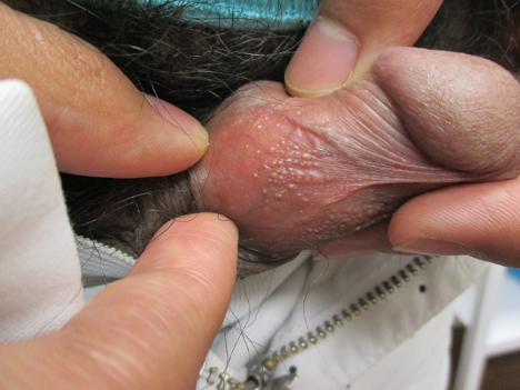
Figure 2. Fordyce spots occur less commonly on the penis.
Acrochordons
Acrochordons, also known as skin tags, molluscum pendulum, or fibroepithelial polyps, are the most common fibrous lesion of the skin. Histologically, the lesion has a flattened epithelium or a folded epithelium, which may be acanthotic and hyperpigmented. The stroma has loose connective tissue with dilated blood vessels. Skin appendages and nerves are usually absent.
Acrochordons occur in approximately 46% of the general population; the incidence increases with age.16 This condition is often associated with obesity and insulin resistance.17-19
Typically, acrochordons present as soft, flesh-colored to dark brown, sessile or pedunculated skin growths with a smooth contour.20,21 Occasionally, the lesions may be hyperkeratotic or have a warty appearance.16 Most acrochordons are 2 to 5 mm in diameter, although they are often larger in the groin.21 Acrochordons are usually asymptomatic. Occasionally, they can be pruritic or painful when inflamed.16 Acrochordons can appear on almost any part of the body but are most frequently seen on the neck (Figure 3) and intertriginous areas (Figures 4 and 5).20-22 Acrochordons on the neck is a remarkable feature of tuberous sclerosis complex.22

Figure 3. Acrochordons can appear on almost any part of the body but are most frequently seen on the neck.
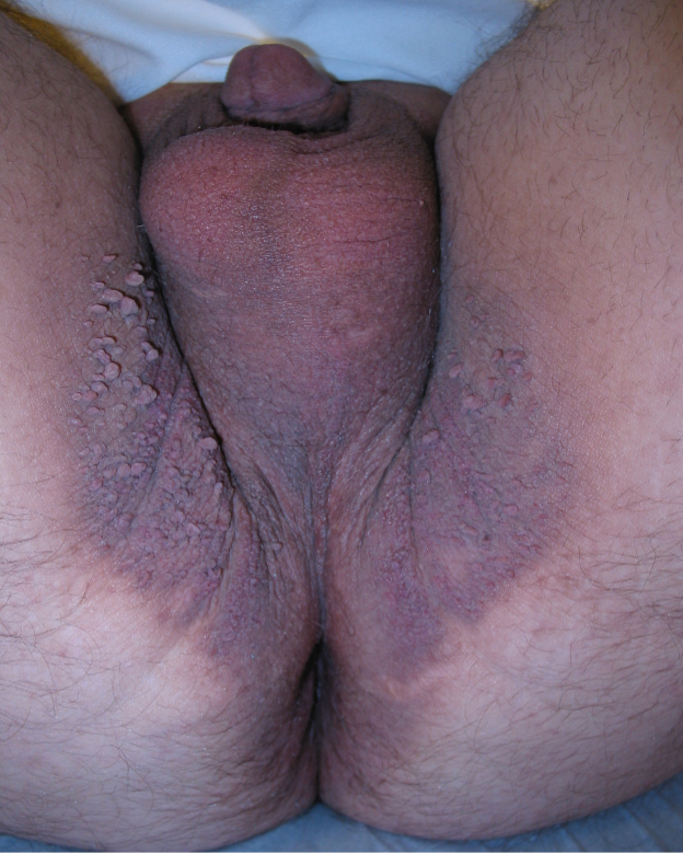
Figure 4. Acrochordons can appear in almost any part of the body but are most frequently seen on the intertriginous areas.

Figure 5. Acrochordons can appear in almost any part of the body but are most frequently seen on the intertriginous areas.
REFERENCES
1. Mutizwa MM, Berk DR. Dichotomous long-term response to isotretinoin in two patients with Fordyce spots. Pediatr Dermatol. 2014;31(1):73-75. https://doi.org/10.1111/j.1525-1470.2012.01749.x
2. Baeder FM, Pelino JE, de Almeida ER, Duarte DA, Santos MT. High-power diode laser use on Fordyce granule excision: a case report. J Cosmet Dermatol. 2010;9(4):321-324. https://doi.org/10.1111/j.1473-2165.2010.00531.x
3. Chern PL, Arpey CJ. Fordyce spots of the lip responding to electrodesiccation and curettage. Dermatol Surg. 2008;34(7):960-962. https://doi.org/10.1111/j.1524-4725.2008.34187.x
4. Radhakrishnan S, Agarwal DC. Fordyce spots masquerading as penile warts. Med J Armed Forces India. 2016;72(4):384-385. https://doi.org/10.1016/j.mjafi.2015.12.008
5. Ocampo-Candiani J, Villarreal-Rodríguez A, Quiñones-Fernández AG, Herz-Ruelas ME, Ruíz-Esparza J. Treatment of Fordyce spots with CO2 laser. Dermatol Surg. 2003;29(8):869-871. https://doi.org/10.1046/j.1524-4725.2003.29236.x
6. Mansur AT, Aydingoz IE. Unilateral buccal Fordyce spots with ipsilateral facial paralysis: a sign of neuro-sebaceous connection? Acta Derm Venereol. 2012;92(2):177-178. https://doi.org/10.2340/00015555-1218
7. Arun Babu T, Vijayadevagaran V, Carounanidy U. Congenital intraoral Fordyce spots. Arch Dis Child Fetal Neonatal Ed. 2016;101(3):F252. https://doi.org/10.1136/archdischild-2015-309986
8. Leung AKC, Barankin B. Fordyce spots. Clin Case Rep Rev. 2015;1(6):121-122. doi:10.15761/CCRR.1000140
9. Plotner AN, Brodell RT. Treatment of Fordyce spots with bichloracetic acid. Dermatol Surg. 2008;34(3):397-399. https://doi.org/10.1111/j.1524-4725.2007.34078.x
10. Fernandez-Flores A, Rodríguez Peralto JL. Mismatch repair protein expression in fordyce granules. Appl Immunohistochem Mol Morphol. 2017;25(3):209-212. https://doi.org/10.1097/pai.0000000000000339
11. Ahn GR, Park SJ, Lee CK, Kim BJ. A case of successful treatment of Fordyce spots with a single insulated microneedle radiofrequency device. Dermatol Ther. 2019;32(5):e13026. https://doi.org/10.1111/dth.13026
12. Jakhar D, Kaur I. Mucoscopy of Fordyce's Spots on Lips. Indian Dermatol Online J. 2019;10(4):498-499. https://doi.org/10.4103/idoj.idoj_185_18
13. Rane V, Read T. Penile appearance, lumps and bumps. Aust Fam Physician. 2013;42(5):270-274. https://www.racgp.org.au/afp/2013/may/penile-appearance/
14. Pallua N, Stromps JP. Micro-punch technique for treatment of Fordyce spots: a surgical approach for an unpleasant condition of the male genital. J Plast Reconstr Aesthet Surg. 2013;66(1):e8-e11. https://doi.org/10.1016/j.bjps.2012.08.039
15. Gaballah KY, Rahimi I. Can presence of oral Fordyce's granules serve as a marker for hyperlipidemia? Dent Res J (Isfahan). 2014;11(5):553-558. https://www.ncbi.nlm.nih.gov/pubmed/25426145
16. Belgam Syed SY, Lipoff JB, Chatterjee K. Acrochordon. In: StatPearls. Treasure Island (FL): StatPearls Publishing; August 10, 2020. http://www.ncbi.nlm.nih.gov/books/nbk448169/
17. Marchand L, Gaimard M, Luyton C. All about skin manifestations of insulin resistance and type 2 diabetes: acanthosis nigricans and acrochordons. Postgrad Med J. 2020;96(1134):237. https://doi.org/10.1136/postgradmedj-2019-136834
18. Ragunatha S, Anitha B, Inamadar AC, Palit A, Devarmani SS. Cutaneous disorders in 500 diabetic patients attending diabetic clinic. Indian J Dermatol. 2011;56(2):160-164. https://doi.org/10.4103/0019-5154.80409
19. Shah R, Jindal A, Patel N. Acrochordons as a cutaneous sign of metabolic syndrome: a case-control study. Ann Med Health Sci Res. 2014;4(2):202-205. https://doi.org/10.4103/2141-9248.129040
20. Bahce ZS, Akbulut S, Sogutcu N, Oztas T. Giant acrochordon arising from the thigh. J Coll Physicians Surg Pak. 2015;25(11):839-840. https://doi.org/11.2015/jcpsp.839840
21. Ma H, Xia Y, Yin S, Lai W. Giant skin tag on the labium majorum. Int J Womens Dermatol. 2015;1(4):175-176. https://doi.org/10.1016/j.ijwd.2015.09.003
22. Baykal C. Acrochordons on the neck; a remarkable clinical feature of tuberous sclerosis showing different patterns. J Eur Acad Dermatol Venereol. 2018;32(4):e146-e147. https://doi.org/10.1111/jdv.14636