
Peer Reviewed
Agminated Blue Nevus Arising in a Nevus Spilus
Authors:
Margaret C. Green, DO, MSc
Department of Dermatology, Naval Medical Center San Diego, California
Jordyn G. Shah, DO
Naval Medical Center San Diego, California
Daryl J. Sulit, MD
Department of Dermatology, Naval Medical Center San Diego, California, and Assistant Professor of Dermatology at the Uniformed Services University of the Health Sciences School of Medicine in Bethesda, Maryland
Citation:
Green MC, Shah JG, Sulit DJ. Agminated blue nevus arising in a nevus spilus. Consultant. 2019;59(8):239-241.
A 34-year-old Hispanic woman with Fitzpatrick skin type III presented to a dermatology clinic with what she described as a changing birthmark.
She reported having a light brown patch on her left forearm with multiple darker-brown 2- to 4-mm macules that had become studded with multiple blue 3- to 4-mm papules within the past few years (Figures 1-3). The lesion was asymptomatic and had not changed in size.

Figure 1. Clinical photograph; site selected for biopsy encircled.

Figure 2. Nevus spilus background studded with multiple blue nevi with intervening pigment between them.
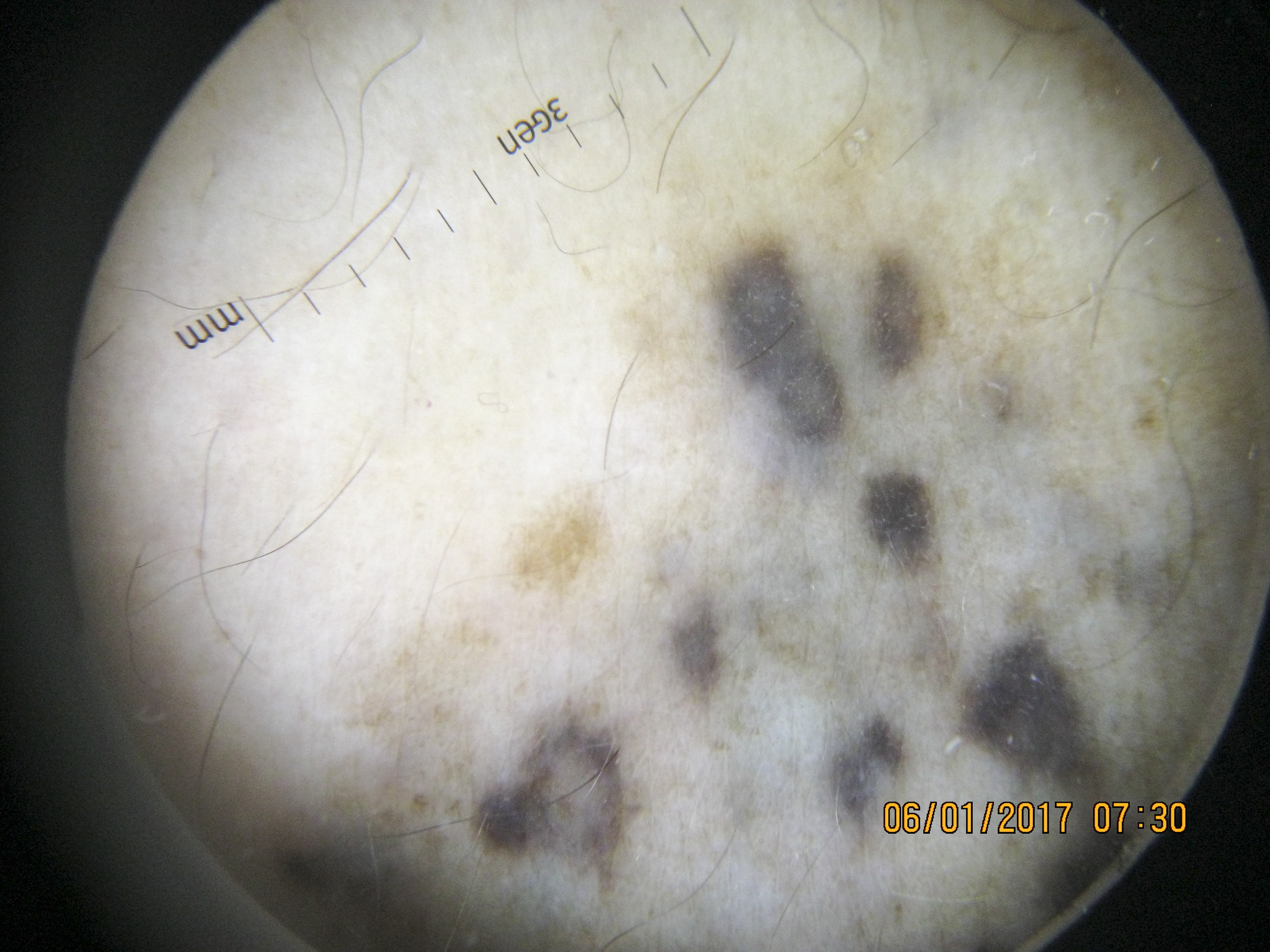
Figure 3. Dermoscopy photograph demonstrating a lentiginous component and homogenous blue papules.
On examination, a 3 × 2-cm light brown to gray patch studded with darker brown macules and blue papules was present on her left forearm. A punch biopsy of a representative “new” papule was obtained (Figure 1). The pathology report showed junctional melanocytic proliferation in the epidermis (consistent with nevus spilus) and spindled cells associated with melanophages and sclerotic collagen in the superficial to deep reticular dermis (consistent with common blue nevus) (Figures 4-6). Together, these findings were consistent with an agminated blue nevus within a nevus spilus.

Figure 4. Junctional melanocytic proliferation in the epidermis (consistent with junctional nevus portion of nevus spilus) and spindled cells associated with melanophages and sclerotic collagen in the superficial to deep reticular dermis (consistent with common blue nevus) (hematoxylin-eosin, original magnification ×4).

Figure 5. High-power view of junctional nevus portion of nevus spilus (hematoxylin-eosin, original magnification ×40).

Figure 6. High-power view of common blue nevus in reticular dermis (hematoxylin-eosin, original magnification ×40).
After discussion with the patient about the diagnosis and prognosis, the follow-up plan was once-a-year skin examinations at the dermatology clinic. However, if the lesion changed color or significantly increased in size, then an excisional biopsy or incisional biopsy would be considered to rule out a malignant transformation.
DISCUSSION
Nevus spilus is a melanocytic lesion made up of collection of multiple dark-brown macules to papules, occurring within a lighter background patch.1 Although nevus spilus is considered benign, it has been proposed that the lesion may create an environment that promotes the development of other nevi2,3 and rarely cutaneous malignancies.2,4,5 Nevus spilus often presents at birth or during childhood, but the development of other types of nevi within the nevus spilus often occurs years later.1,3,4,6
The term agminated means “clustered.” Concerning the variant of plaque-type blue nevus known as an agminated blue nevus, the clinical presentation usually consists of a faint blue-gray patch background flecked with darker blue-black macules or papules. Histologically, the papular component usually represents a common blue nevus, while the patch component represents dermal melanocytosis similar to that of nevus of Ota or mongolian spot.7 Histologically, both components are comprised of melanocytes in the reticular dermis with dendritic morphology, but the common blue nevus has a much denser collection.7
The background patch component of nevus spilus often shows features of lentigo simplex with elongated rete ridges, an increased number of basal layer melanocytes, and increased melanin in the epidermis and in dermal melanophages.7 The darker macules or papules are most commonly benign junctional, compound, or even dermal nevi.7
Nevus spilus is a fairly rare entity, with incidence estimated to be 0.2% to 2.3% among the general population.5 The malignant potential for development of melanoma within a nevus spilus is currently unknown, but Brito and colleagues reported a risk of 0.13% to 0.20%, with greater risk in a nevus spilus larger than 4 cm (medium to giant) and those present from birth or early childhood.5
Development of malignant melanoma is also reported within blue nevi. Borgenvik and colleagues recently reviewed the literature and reported 48 cases of melanoma arising within a previously benign blue nevus.8 Most of these cases (26) developed within a cellular blue nevus, but 3 cases of melanoma developed within a plaque-type blue nevus.8
A total of 13 cases of agminated blue nevus arising within a nevus spilus with histopathologic verification were identified in a PubMed literature search including English, French, and German language publications.1-6,8-16 However, Ishibashi and colleagues suggested that this entity may be underreported and more common in the Japanese population.14 Additionally, 2 cases described histopathologically as a combination of pigmented cells in the epidermis and blue nevus cells in the dermis were first described by Montgomery and Kahler in 1939,15 and this combined entity was first classified by Kawamura in 1950 as a type 2 atypical blue nevus.16 However, it is difficult to determine whether these additional cases represent the combination of nevus spilus and agminated blue nevus, given the difference in terminology and lack of clinical photographs. Most of the 13 cases had a similar clinical presentation to our patient—a long-standing or congenital nevus spilus that later developed blue nevi.
To our knowledge, there has been 1 reported case of a malignant melanoma arising within a congenital nevus spilus, which also contained acquired, benign blue nevi,4 and 1 reported case of a superficial basal cell carcinoma arising within a similar lesion.2
Since an agminated blue nevus within a nevus spilus is a rarely reported entity, its prognosis is uncertain, making patient counseling challenging. It is unknown whether the risk of malignant transformation is higher or lower compared with separate nevus spilus or classic blue nevus lesions. It is assumed that the risk of malignant transformation is low, but it is possible that melanoma can develop within either the nevus spilus component or the blue nevus component separately.
For our patient, clinical photographs and measurements were taken at the time of her initial visit, recognition of concerning changes was discussed, and annual follow-up examinations were recommended.
REFERENCES:
- Ayala D, Ramón MD, Cabezas M, Jordá E. Nevus spilus associated with agminated blue nevus: a rare combination. Actas Dermosifiliogr. 2016;107(7):614-616.
- Betti R, Inselvini E, Crosti C. Blue nevi and basal cell carcinoma within a speckled lentiginous nevus. J Am Acad Dermatol. 1999;41(6):1039-1041.
- Marchesi L, Naldi L, Parma A, Locati F, Cainelli T. Agminate blue nevus combined with lentigo: a variant of speckled lentiginous nevus? Am J Dermatopathol. 1993;15(2):162-165.
- Yoneyama K, Kamada N, Mizoguchi M, Utani A, Kobayashi T, Shinkai H. Malignant melanoma and acquired dermal melanocytosis on congenital nevus spilus. J Dermatol. 2005 Jun;32(6):454-458.
- Brito MHTS, Dionísio CSNM, Fernandes CMBM, Ferreira JCM, Rosa MJMPMC, Garcia MMAPS. Synchronous melanomas arising within nevus spilus. An Bras Dermatol. 2017;92(1):107-109.
- Simonetti V, Grenzi L, Piana S, Albertini G, Longo C. Agminated blue nevus combined with nevus spilus: an uncommon association. Int J Dermatol. 2015;54(2):215-21
- Luzur B, Bastian BC, Calonje E. Melanocytic nevi. In: Calonje E, Brenn T, Lazar A, McKee PH, eds. McKee’s Pathology of the Skin with Clinical Correlations. Vol 2. 4th ed. Philadelphia, PA: Elsevier Saunders; 2012:chap 25.
- Borgenvik TL, Karlsvik TM, Ray S, Fawzy M, James N. Blue nevus-like and blue nevus-associated melanoma: a comprehensive review of the literature. ANZ J Surg. 2017;87(5):345-349.
- Kiene P, Brodersen JP, Fölster-Holst R. “Blue” variant of naevus spilus [in German]. Hautarzt. 1995;46(5):349-351.
- Toppe F, Haas N. Giant nevus spilus with blue nevus. Apropos of a case [in French]. Ann Dermatol Venereol. 1988;115(6-7):703-707.
- Misago N, Narisawa Y, Kohda H. A combination of speckled lentiginous nevus with patch-type blue nevus. J Dermatol. 1993;20(10):643-647.
- Park YM, Kang H, Cho BK. Plaque-type blue nevus combined with nevus spilus and smooth muscle hyperplasia. Int J Dermatol. 1999;38(10):775-777.
- Cox NH, Malcolm A, Long ED. Superficial spreading melanoma and blue naevus within naevus spilus—ultrastructural assessment of giant pigment granules. Dermatology. 1997;194(3):213-216.
- Ishibashi A, Kimura K, Kukita A. Plaque-type blue nevus combined with lentigo (nevus spilus). J Cutan Pathol. 1990;17(4):241-245.
- Montgomery H, Kahler JE. The blue nevus (Jadassohn-Tieche): its distinction from ordinary moles and malignant melanomas. Am J Cancer. 1939;36(4):527-539.
- Kawamura T. Atypical blue nevus; report of a case. Arch Derm Syphilol. 1950;62(3):395-399.