
Peer Reviewed
Caught in the Act: Thrombus Straddling a Patent Foramen Ovale
AUTHORS:
Ramtej Atluri, MBBS1 • Anuradha Kolluru, MD2
AFFILIATIONS:
1NRI Medical College, Guntur, Andhra Pradesh, India
2Decatur Memorial Hospital, Decatur, Illinois
CITATION:
Atluri R, Kolluru A. Caught in the act: thrombus straddling a patent foramen ovale. Consultant. 2020;60(6):e6. doi:10.25270/con.2020.04.00010
Received December 10, 2019. Accepted March 31, 2020.
DISCLOSURES:
The authors report no relevant financial relationships.
CORRESPONDENCE:
Anuradha Kolluru, MD, Illinois Heart Specialists, 2300 N Edward St, Decatur, IL 62526 (akolluru2012@gmail.com)
A 74-year-old, morbidly obese, sedentary man presented with a 2-day history of acute-onset dyspnea. At presentation, he was significantly hypoxic and tachypneic, and physical examination revealed bilateral lower extremity edema.
Further workup with Duplex ultrasonography revealed bilateral deep venous thrombosis. Computed tomography angiography of the chest showed thrombi bilaterally in the pulmonary arteries, the atria, and the right ventricle, with evidence of right ventricular strain. Transthoracic echocardiography was limited due to the patient’s body habitus.
Transesophageal echocardiography (TEE) demonstrated thrombi in transit through a patent foramen ovale (PFO) (Figures 1 and 2; Video 1) and showed the presence of thrombi in all 4 chambers of the heart (Figure 3; Video 2).
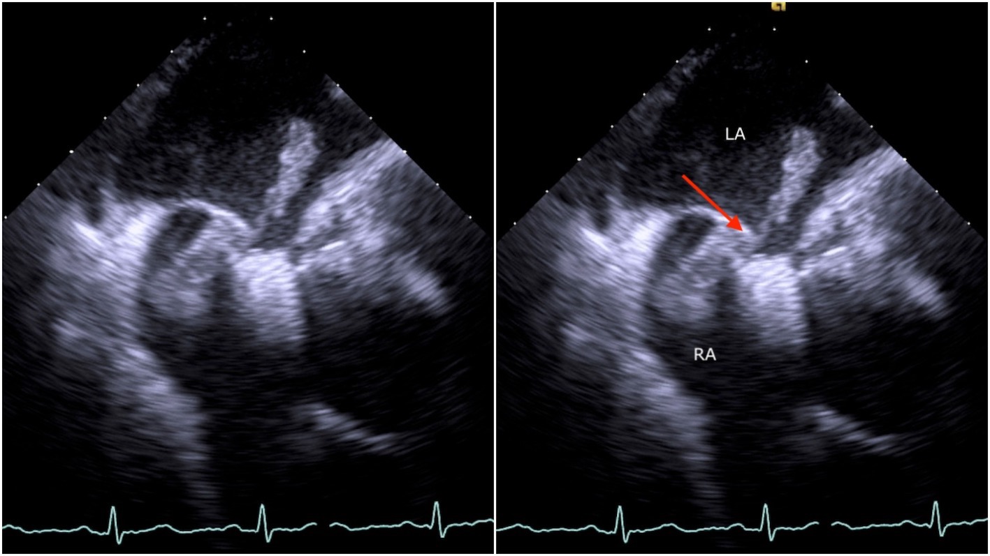
Figure 1. TEE image demonstrating a thrombus traversing a patent foramen ovale (red arrow). Abbreviations: LA, left atrium; RA, right atrium.
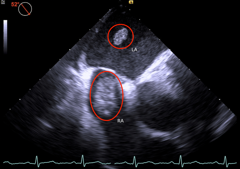
Figure 2. TEE image demonstrating thrombi in both the right and left atria (marked in red). Abbreviations: LA, left atrium; RA, right atrium.
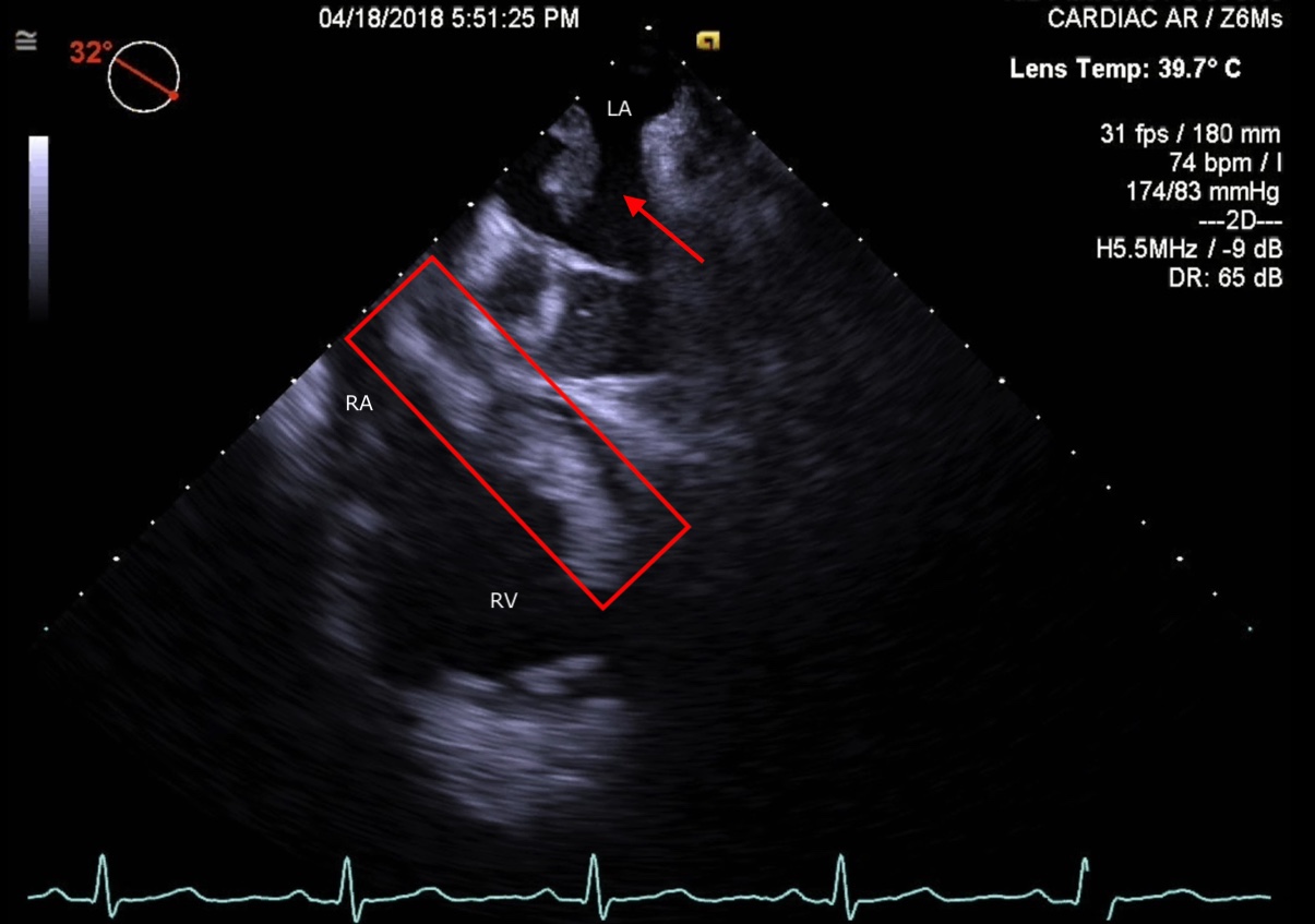
Figure 3. TEE Image depicting thrombus extension into the right ventricle and thrombi in the atria (marked in red). Abbreviations: LA, left atrium; RA, right atrium; RV, right ventricle.
Video 1. TEE video demonstrating a thrombus in transit through a patent foramen ovale.
Video 2. TEE video showing the presence of thrombi in all 4 chambers of the heart.
PFO is found in 20% to 25% of the adult population and is associated with cryptogenic stroke, with paradoxical embolization being the potential mechanism.1,2 Thrombi crossing through a PFO is a well-known occurrence, but it is difficult to visualize because it occurs transiently.
Paradoxical embolization is a well-known mechanism of cryptogenic stroke and systemic embolization in patients with PFO. This is a medical emergency that requires immediate and appropriate management. Anticoagulation, surgical removal of large thrombi (especially in the left heart), and PFO closure are considered as mainstay treatment options. However, given his comorbidities, our patient was managed medically with intravenous thrombolysis and long-term anticoagulation, resulting in a successful outcome. Follow-up TEE revealed complete resolution of thrombi.
It remains unclear how PFO closure compares with systemic anticoagulation in the prevention of recurrent ischemic stroke. PFO closure is of moderate benefit compared with antiplatelet therapy alone in the prevention of recurrent ischemic stroke in adults up to 60 years of age.1,2
REFERENCES:
- Hagen PT, Scholz DG, Edwards WD. Incidence and size of patent foramen ovale during the first 10 decades of life: an autopsy study of 965 normal hearts. Mayo Clin Proc. 1984;59(1):17-20. doi:10.1016/s0025-6196(12)60336-x
- Mojadidi MK, Zaman MO, Elgendy IY, et al. Cryptogenic stroke and patent foramen ovale. J Am Coll Cardiol. 2018;71(9):1035-1043. doi:10.1016/j.jacc.2017.12.059