
Peer Reviewed
Pyoderma Gangrenosum on the Lower Extremity
AFFILIATIONS:
1Medical student, University of Illinois at Chicago-Rockford, Chicago, Illinois
2Dermatology resident, Naval Medical Center, San Diego, California
CITATION:
Brubaker R, Schmiedecke R. Pyoderma gangrenosum on the lower extremity. Consultant. 2022;62(12):e10. doi:10.25270/con.2022.08.000002
Received March 23, 2022. Accepted July 7, 2022. Published online August 2, 2022.
DISCLOSURES:
The authors report no relevant financial relationships.
DISCLAIMER:
The authors report that informed patient consent was obtained for publication of the images used herein.
CORRESPONDENCE:
Riley Brubaker, BS, University of Illinois at Chicago-Rockford, 1601 Parkview Ave, Rockford, IL 61107 (rileybrubaker96@gmail.com)
A 53-year-old woman presented for a dermatology consult for a spreading ulcer on the lateral left calf. The patient thought the lesion was an insect bite and had applied an antibiotic ointment and a bandage. A few days later, the lesion became more painful, with surrounding erythema and drainage (Figure 1).

Figure 1. A 9-cm painful ulcer with granulation tissue, violaceous borders, and surrounding erythema was noted on the left lower extremity.
Over the next week, she visited her primary care physician, an urgent care physician, and the emergency department and was treated for cellulitis with treatment including warm compresses and several courses of antibiotics (cefalexin, 500 mg 4 times a day for 10 days; doxycycline, 100 mg twice daily for 14 days; two doses of vancomycin, 1500 mg; 1 dose of meropenem, 2 g; and 1 dose of clindamycin, 900 mg). A wound culture taken during her initial presentation showed clindamycin- and tetracycline-resistant Streptococcus agalactiae; later blood and tissue cultures were negative for growth. A computed tomography scan of the left lower extremity was performed because of concern for possible necrotizing fasciitis or osteomyelitis.
Physical examination. The lesion measured 9 cm, was exquisitely tender, and consisted of abundant granulation tissue with sparse fibrinous debris, undermined and violaceous borders, and surrounding warmth and erythema. She had a medical history significant for type 2 diabetes and well-controlled ulcerative colitis on 6-mercaptopurine and sulfasalazine.
Treatment and management. Given the pertinent history and physical examination, the condition was diagnosed as pyoderma gangrenosum and was treated with 5 g topical clobetasol 0.05% ointment twice daily under nonadherent dressings. Her pain improved significantly after a single day of treatment. After 3 weeks of treatment, the lesion improved significantly (Figure 2).
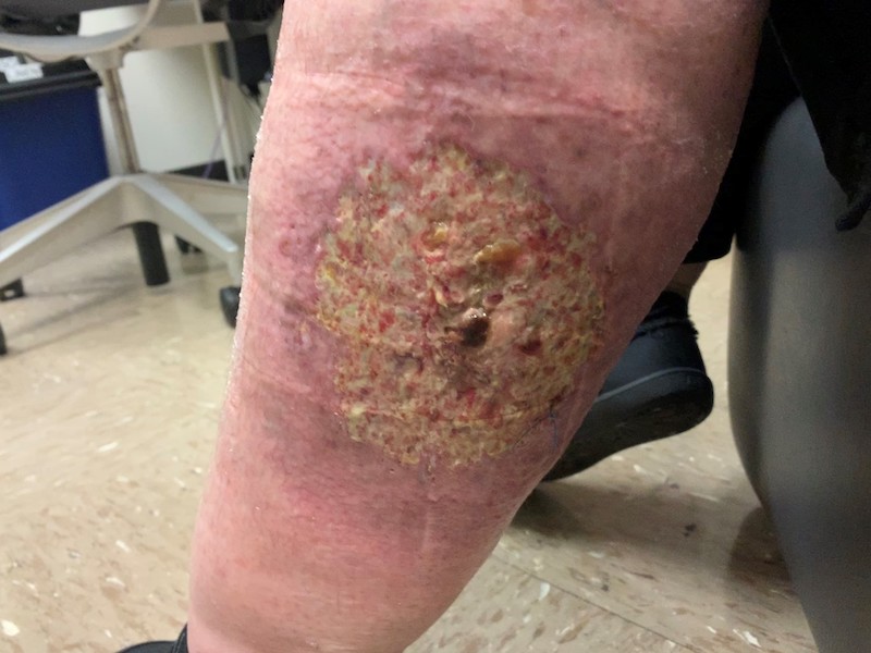
Figure 2. The lesion improved significantly after 3 weeks of treatment with 5 g topical clobetasol 0.05% ointment twice daily under nonadherent dressings.
Discussion. Pyoderma gangrenosum is a self-limiting, neutrophilic dermatosis that most often manifests as a painful, purulent, rapidly growing ulcer with an irregular, violaceous border and peripheral erythema. Ulcerative pyoderma gangrenosum is the most common presentation, but other clinical variants exist.1 The incidence of the disease is reported at 3 to 10 per million persons per year.2 It frequently arises on the lower extremities and trunk, but other sites may also be involved. Pyoderma gangrenosum commonly affects women aged 40 to 60 years, and an associated underlying medical condition is often present, such as inflammatory bowel disease, hematologic disorders, infection, or inflammatory arthritides. Lesions may exhibit pathergy (spread with trauma).1
The clinical presentation is important because pyoderma gangrenosum is a diagnosis of exclusion and no definitive diagnostic serology or histology exists. Arthropod assault (insect bites), infection, and vasculitis are often misdiagnosed for pyoderma gangrenosum because of their commonality. However, a violaceous or undermined border around the lesion can help distinguish pyoderma gangrenosum.2 In addition, if any of the aforementioned underlying conditions are present, then a diagnosis of pyoderma gangrenosum should be strongly considered. Insect bites often get better, not worse, and if worsening does occur, then other systemic symptoms are usually reported.1 Although pyoderma gangrenosum can grow quickly (within days), necrotizing fasciitis spreads very rapidly, with the hallmark symptom of pain at rest and out of proportion to examination. Early diagnosis of pyoderma gangrenosum is crucial because it can significantly reduce patient morbidity potentially caused by unnecessary treatment and interventions as well as disfiguring scarring, psychological consequences, and multiple care visits.2
Treatment of pyoderma gangrenosum depends on severity. Stable or slowly growing lesions can be treated with compresses with occlusive dressing as pyoderma gangrenosum can spontaneously regress. Potent topical corticosteroids, topical dapsone, or topical tacrolimus can be considered for more persistent lesions.1 Rapid progression, extensive disease, or facial involvement may necessitate the use of systemic corticosteroids, cyclosporine, or infliximab. If an underlying associated disease is present, treatment of that disorder is recommended and can lead to improvement of the pyoderma gangrenosum.4 Response to treatment is typically seen in 1 to 2 weeks, but resolution may take months.1 Caution should be given to prolonged use of high-dose, super-potent topical corticosteroids to avoid potential complications, such as elevated blood glucose level, Cushing syndrome, or adrenal suppression.5,6 Limiting the risk of pathergy (eg, by avoiding unnecessary surgical procedures) and good wound care are also vital for resolution.3
Conclusion. A diagnosis of pyoderma gangrenosum is often delayed for multiple reasons, incIuding variable presentation, more common acute or severe cutaneous disorders that mimic pyoderma gangrenosum, and relative rarity of these lesions in primary and acute care settings. First-line treatments include gentle wound care and potent topical corticosteroids with a taper as the lesion improves. When in doubt, consider consulting a dermatologist.
1. James WD, Elston DM, Treat JR, Rosenbach MA, Neuhaus IM. Erythema and urticaria. In: Andrews’ Diseases of the Skin. 13th ed. Elsevier: 2019:140-156.e3.
2. Bisarya K, Azzopardi S, Lye G, Drew PJ. Necrotizing fasciitis versus pyoderma gangrenosum: securing the correct diagnosis! A case report and literature review. Eplasty. 2011;11:e24.
3. Bolognia JL, Schaffer, JV, Duncan KO, Ko CJ. Neutrophilic dermatoses. In: Dermatology Essentials. Elsevier; 2014:199-208.
4. Lebwohl MG, Heymann WR, Coulson IH, Murrell DF. Pyoderma gangrenosum. In: Treatment of Skin Disease: Comprehensive Therapeutic Strategies. 6th ed. 2021;711-715.
5. Nakamura M, Abrouk M, Zhu H, Farahnik B, Koo J, Bhutani T. Update on the systemic risks of superpotent topical steroids. J Drugs Dermatol. 2017;16(7):643-648.
6. Gilbertson EO, Spellman MC, Piacquadio DJ, Mulford MI. Super potent topical corticosteroid use associated with adrenal suppression: Clinical considerations. J Am Acad Dermatol. 1998;38(2):318-321. doi.10.1016/S0190-9622(98)70573-0