
Peer Reviewed
An Atlas of Nail Disorders, Part 10
AUTHORS:
Alexander K. C. Leung, MD1,2 • Benjamin Barankin, MD3 • Kin Fon Leong, MD4
AFFILIATIONS:
1Department of Pediatrics, University of Calgary, Calgary, Alberta, Canada
2Alberta Children’s Hospital, Calgary, Alberta, Canada
3Toronto Dermatology Centre, Toronto, Ontario, Canada
4Pediatric Institute, Kuala Lumpur General Hospital, Kuala Lumpur, Malaysia
CITATION:
Leung AKC, Barankin B, Leong KF. An atlas of nail disorders, part 10. Consultant. 2020;60(8):19-21. doi:10.25270/con.2020.08.00003
DISCLOSURES:
The authors report no relevant financial relationships.
CORRESPONDENCE:
Alexander K. C. Leung, MD, #200, 233 16th Ave NW, Calgary, AB T2M 0H5, Canada (aleung@ucalgary.ca)
EDITOR’S NOTE:
This article is part 10 of a 15-part series of Photo Essays describing and differentiating conditions affecting the nails. Parts 11 through 15 will be published in upcoming issues of Consultant. To access previously published articles in the series, visit the Consultant archive at www.Consultant360.com and click the “Journals” tab.
Psoriatic Nails
Psoriasis, a chronic inflammatory immune-mediated disease involving the skin, the nails, and the musculoskeletal structures, affects 1% to 3% of the world population.1,2 Approximately 25% of persons with psoriasis develop the disease before 20 years of age.3 Plaque psoriasis or psoriasis vulgaris, the most common variant, is characterized by sharply demarcated erythematous plaques with adherent silvery micaceous scales (Figure 1).3 Removal of the scales results in fine punctate bleeding, which is referred to as the Auspitz sign.3 The lesions are usually symmetrically distributed and can be pruritic. Typical sites include the knees (Figure 2), elbows, and lower back. Involvement of the scalp (Figure 3), face, and the intertriginous and diaper areas is more common in infants and young children.3 Mucosal involvement is unusual. Other variants include guttate psoriasis, pustular psoriasis, erythrodermic psoriasis, and flexural (or inverse) psoriasis.3 Seronegative inflammatory arthritis develops in approximately 5% to 30% of persons with psoriasis.3 Psoriatic arthritis can precede, coincide with, or follow (most commonly) the development of the skin lesions.
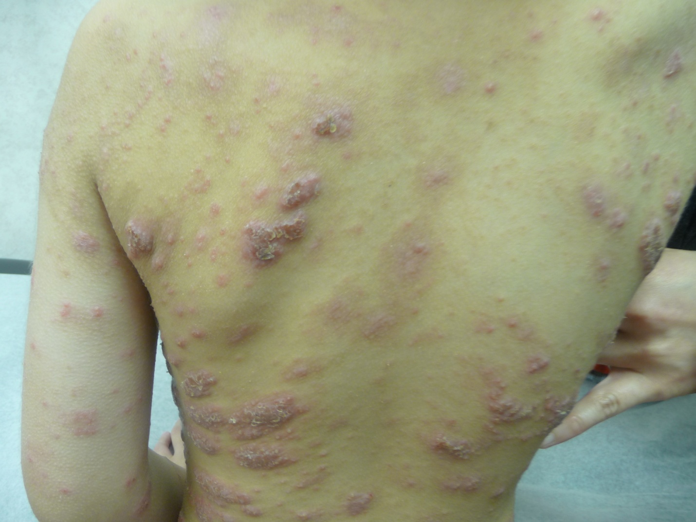
Figure 1

Figure 2
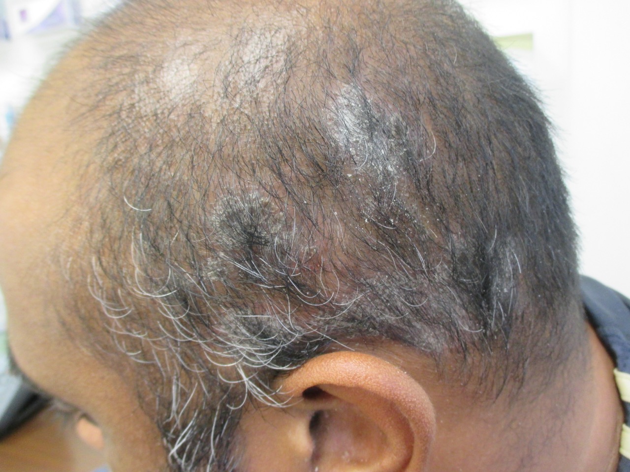
Figure 3
Up to 90% of patients with psoriasis eventually develop nail involvement, especially those with psoriatic arthritis.2,4-6 Nail psoriasis is slightly more common in males than females.7 All or a few fingernails or toenails can be involved.2,6 Generally, fingernails are more frequently affected than toenails.4,7 Nail psoriasis is less common in children, where the prevalence is 7% to 32%.2,8,9 The prevalence increases with the age of the child and is more common in boys.9 Approximately 5% of patients have isolated nail psoriasis without other cutaneous manifestations.6,7,10 Nail involvement precedes the skin lesions in approximately 4% of patients.11
Nail involvement includes nail pitting (Figure 4), trachyonychia, onychauxis, onychorrhexis (“brittle nails”), leukonychia (Figure 5), onycholysis (Figure 6), Beau lines, onychomadesis, oil drop discoloration, red spots (salmon patches) in the lunula (mottled lunula), dyschromias, longitudinal ridges, subungual hyperkeratosis (Figure 7), splinter hemorrhage (Figure 8), nail plate crumbling (Figure 9), or a combination thereof.1,2,5-7,12-17 Nail plate crumbling may progress to total nail matrix destruction after a prolonged period of disease activity.15 By far, pitting is the most common manifestation of nail psoriasis and occurs in approximately 70% of patients with nail psoriasis.7,10,18 It has been proposed that 20 to 60 pits are suggestive of psoriasis whereas greater than 60 pits are highly specific for psoriasis.18 Other less common manifestations include acropustulosis, subacute or chronic paronychia, and psoriatic onycho-pachydermo-periostitis.7 Studies have shown that nail psoriasis is an independent prognostic factor for the development of psoriatic arthritis.5,6,19

Figure 4

Figure 5
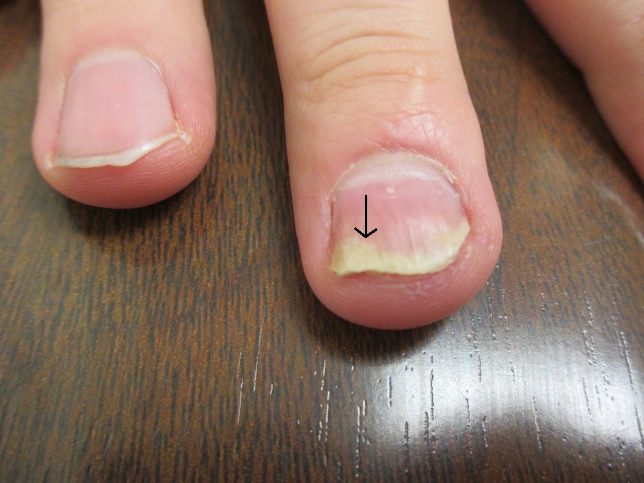
Figure 6

Figure 7

Figure 8

Figure 9
Dermoscopy can be helpful in the diagnosis when the clinical features are not typical. Dermoscopic features suggestive of nail psoriasis include coarse, deep, large pits that are irregular in shape, size, and distribution; diffuse scales, especially in the proximal part of the nail; slightly dented, erythematous margin surrounding the distal edge of the detachment; purplish black or reddish brown longitudinal lines on the nail bed; salmon patches; and irregularly distributed, dilated, and tortuous capillaries at the hyponychium or the proximal nail fold.2,4,19,20
Nail psoriasis can be cosmetically unsightly and socially embarrassing.2,4 The condition can cause undue emotional distress and can have an adverse effect on quality of life.4,6,7,21 Other complications of psoriasis include increased risk of onychomycosis and nonmelanoma skin cancer.6,21 Psoriatic onychodystrophy can clinically mimic onychomycosis, and differentiation of the two conditions can be difficult, especially in patients with isolated nail psoriasis and when only the toenails are affected.2,4,22 After all, the two conditions may coexist.22 It is interesting to note that onychomycosis could exacerbate psoriatic onychodystrophy through the Koebner phenomenon.23 A systematic review of 10 studies showed an average prevalence of 18% of onychomycosis in psoriatic patients.24 A recent study of 159 patients (mean age, 44 years) with psoriasis found that one-third of these patients had nail involvement with concomitant onychomycosis.25 Because the treatment of the psoriatic onychodystrophy and onychomycosis are different, it is important to rule out onychomycosis in patients with psoriasis as a cause of onychodystrophy and in patients with psoriatic onychodystrophy. In case of doubt, potassium hydroxide examination of nail scrapings/clippings and fungal culture to demonstrate dermatophytic (the most common cause) hyphae and arthrospores are the best means of confirming the diagnosis.11,25
REFERENCES:
- Al-Mutairi N, Manchanda Y, Nour-Eldin O. Nail changes in childhood psoriasis: a study from Kuwait. Pediatr Dermatol. 2007;24(1):7-10. doi:10.1111/j.1525-1470.2007.00324.x
- Bardazzi F, Starace M, Bruni F, Magnano M, Piraccini BM, Alessandrini A. Nail psoriasis: an updated review and expert opinion on available treatments, including biologics. Acta Derm Venereol. 2019;99(6):516-523. doi:10.2340/00015555-3098
- Leung AKC, Robson WLM. Psoriasis. In: Lang F, ed. Encyclopedia of Molecular Mechanisms of Disease. Springer; 2009:1750-1751. doi:10.1007/978-3-540-29676-8_1489
- Haneke E. Nail psoriasis: clinical features, pathogenesis, differential diagnoses, and management. Psoriasis (Auckl). 2017;7:51-63. doi:10.2147/PTT.S126281
- Kaul S, Singal A, Grover C, Sharma S. Clinical and histological spectrum of nail psoriasis: a cross-sectional study. J Cutan Pathol. 2018;45(11):824-830. doi:10.1111/cup.13334
- Rigopoulos D, Papanagiotou V, Daniel R III, Piraccini BM. Onychomycosis in patients with nail psoriasis: a point to point discussion. Mycoses. 2017;60(1):6-10. doi:10.1111/myc.12542
- Dogra A, Arora AK. Nail psoriasis: the journey so far. Indian J Dermatol. 2014;59(4):319-333. doi:10.4103/0019-5154.135470
- Piraccini BM, Triantafyllopoulou I, Prevezas C, et al. Nail psoriasis in children: common or uncommon? Results from a 10-year double-center study. Skin Appendage Disord. 2015;1(1):43-48. doi:10.1159/000377709
- Pourchot D, Bodemer C, Phan A, et al. Nail psoriasis: a systematic evaluation in 313 children with psoriasis. Pediatr Dermatol. 2017;34(1):58-63. doi:10.1111/pde.13028
- Tan EST, Chong W-S, Tey HL. Nail psoriasis: a review. Am J Clin Dermatol. 2012;13(6):375-388. doi:10.2165/11597000-000000000-00000
- Leung AKC, Leung AAM, Barankin B. Psoriasis with onychodystrophy in a 7-year-old boy. Int J Dermatol Clin Res. 2015;1(2):16-17. doi:10.17352/2455-8605.000007
- Fassio A, Giovannini I, Idolazzi L, Zabotti A, Iagnocco A, Sakellariou G. Nail ultrasonography for psoriatic arthritis and psoriasis patients: a systematic literature review. Clin Rheumatol. 2020;39(5):1391-1404. doi:10.1007/s10067-019-04748-2
- Leung AKC, Leong KF, Barankin B. Trachyonychia. J Pediatr. 2020;216:239-239.e1. doi:10.1016/j.jpeds.2019.08.034
- Nieradko-Iwanicka B. Nail psoriasis — what a rheumatologist should know about. Reumatologia. 2017;55(1):44-47. doi:10.5114/reum.2017.66687
- Schons KRR, Knob CF, Murussi N, Beber AAC, Neumaier W, Monticielo OA. Nail psoriasis: a review of the literature. An Bras Dermatol. 2014;89(2):312-317. doi:10.1590/abd1806-4841.20142633
- Schons KRR, Beber AAC, Beck MO, Monticielo OA. Nail involvement in adult patients with plaque-type psoriasis: prevalence and clinical features. An Bras Dermatol. 2015;90(3):314-319. doi:10.1590/abd1806-4841.20153736
- Uber M, Abagge KT, Robl R, Carvalho VO, Marinoni LP. Nail changes in psoriatic children. Indian J Dermatol Venereol Leprol. 2016;82(3):314-316. doi:10.4103/0378-6323.174380
- Chu DH, Rubin AI. Diagnosis and management of nail disorders in children. Pediatr Clin North Am. 2014;61(2):293-308. doi:10.1016/j.pcl.2013.11.005
- Yorulmaz A, Artuz F. A study of dermoscopic features of nail psoriasis. Postepy Dermatol Alergol. 2017;34(1):28-35. doi:10.5114/ada.2017.65618
- Golińska J, Sar-Pomian M, Rudnicka L. Dermoscopic features of psoriasis of the skin, scalp and nails—a systematic review. J Eur Acad Dermatol Venereol. 2019;33(4):648-660. doi:10.1111/jdv.15344
- Hoy NY, Leung AKC, Metelitsa AI, Adams S. New concepts in median nail dystrophy, onychomycosis, and hand, foot, and mouth disease nail pathology. ISRN Dermatol. 2012;2012:680163. doi:10.5402/2012/680163
- Natarajan V, Nath AK, Thappa DM, Singh R, Verma SK. Coexistence of onychomycosis in psoriatic nails: a descriptive study. Indian J Dermatol Venereol Leprol. 2010;76(6):723. doi:10.4103/0378-6323.72468
- Rigopoulos D, Baran R, Chiheb S, et al. Recommendations for the definition, evaluation, and treatment of nail psoriasis in adult patients with no or mild skin psoriasis: a dermatologist and nail expert group consensus. J Am Acad Dermatol. 2019;81(1):228-240. doi:10.1016/j.jaad.2019.01.072
- Klaassen KMG, Dulak MG, van de Kerkhof PCM, Pasch MC. The prevalence of onychomycosis in psoriatic patients: a systematic review. J Eur Acad Dermatol Venereol. 2014;28(5):533-541. doi:10.1111/jdv.12239
- Tabassum S, Rahman A, Awan S, et al. Factors associated with onychomycosis in nail psoriasis: a multicenter study in Pakistan. Int J Dermatol. 2019;58(6):672-678. doi:10.1111/ijd.14364